Shiba Shintaro, Parajuli Raj Kumar, Sakai Makoto, Oike Takahiro, Ohno Tatsuya, Nakano Takashi
Department of Radiation Oncology, Gunma University Graduate School of Medicine, Maebashi, Japan.
Gunma University Heavy Ion Medical Center, Maebashi, Japan.
Front Oncol. 2020 May 19;10:635. doi: 10.3389/fonc.2020.00635. eCollection 2020.
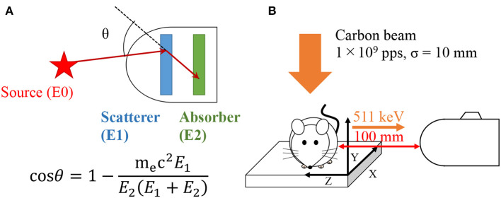
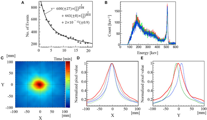
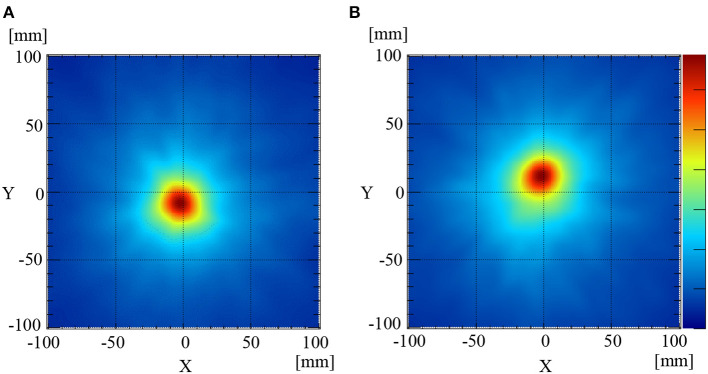
The application of annihilation gamma-ray monitoring to the adaptive therapy of carbon ion radiotherapy (C-ion RT) requires identification of the peak intensity position and confirmation of activated elements with annihilation gamma-rays generated at the C-ion-irradiated site from those transported to unirradiated sites. Real-time monitoring of C-ion-induced annihilation gamma-rays was implemented using a Compton camera in a mouse model. An adult C57BL/6 mouse was anesthetized, and C-ion beams were directed into the abdomen at 1 × 10 particles/s for 20 s. The 511 keV annihilation gamma-rays, generated by the interaction between the irradiated C-ion beam and the target mouse, were detected using a silicon/cadmium telluride (Si/CdTe) Compton camera for 20 min immediately after irradiation. The irradiated site and the peak intensity position of 511 keV gamma emissions due to C-ion beam irradiation on a mouse were observed at the abdomen of the mouse by developing Compton images. Moreover, the positron emitter transport was observed by evaluating the range of gamma-ray emission after the C-ion beam irradiation on the mouse. Our data suggest that by confirming the peak intensity and beam range of C-ion RT with Si/CdTe-based Compton camera, it would be possible to reduce the intra-fractional and inter-fractional dose distribution degradation. Therefore, the results of this study would contribute to the future development of adaptive therapy with C-ion RT for humans.
将湮没伽马射线监测应用于碳离子放射治疗(C离子放疗)的自适应治疗,需要识别峰值强度位置,并确认在C离子照射部位产生的湮没伽马射线与传输到未照射部位的湮没伽马射线中活化元素的情况。在小鼠模型中使用康普顿相机对C离子诱导的湮没伽马射线进行实时监测。将一只成年C57BL/6小鼠麻醉,以每秒1×10个粒子的剂量向其腹部照射C离子束,持续20秒。在照射后立即使用硅/碲化镉(Si/CdTe)康普顿相机对照射的C离子束与目标小鼠相互作用产生的511 keV湮没伽马射线进行20分钟的检测。通过生成康普顿图像,在小鼠腹部观察小鼠C离子束照射导致的511 keV伽马发射的照射部位和峰值强度位置。此外,通过评估小鼠C离子束照射后伽马射线发射范围来观察正电子发射体的传输情况。我们的数据表明,通过使用基于Si/CdTe的康普顿相机确认C离子放疗的峰值强度和束流范围,有可能减少分次内和分次间的剂量分布退化。因此,本研究结果将有助于未来人类C离子放疗自适应治疗的发展。