Parasitology and Mycology Laboratory, Université Cheikh Anta Diop, Dakar, Senegal.
National Malaria Elimination Programme/Epidemiology Division, Department of Public Health, Federal Ministry of Health, Abuja, Nigeria.
Malar J. 2018 Nov 28;17(1):439. doi: 10.1186/s12936-018-2588-7.
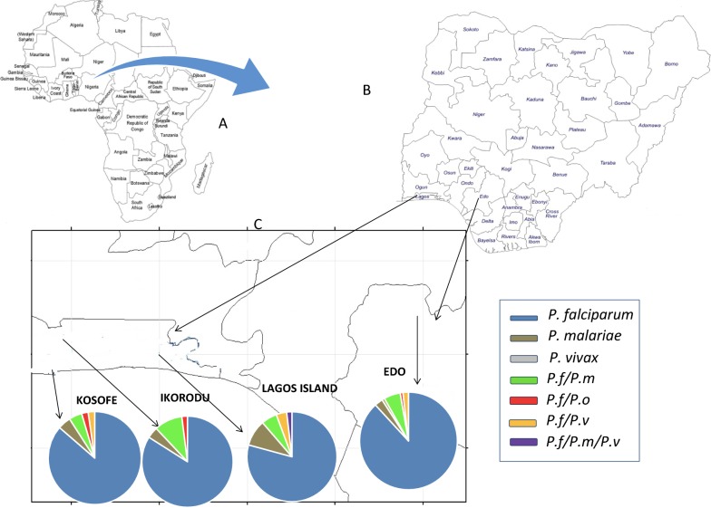
Malaria in Nigeria is principally due to Plasmodium falciparum and, to a lesser extent to Plasmodium malariae and Plasmodium ovale. Plasmodium vivax is thought to be absent in Nigeria in particular and sub-Saharan Africa in general, due to the near fixation of the Duffy negative gene in this population. Nevertheless, there are frequent reports of P. vivax infection in Duffy negative individuals in the sub-region, including reports from two countries sharing border with Nigeria to the west (Republic of Benin) and east (Cameroon). Additionally, there were two cases of microscopic vivax-like malaria from Nigerian indigenous population. Hence molecular surveillance of the circulating Plasmodium species in two states (Lagos and Edo) of southwestern Nigeria was carried out.
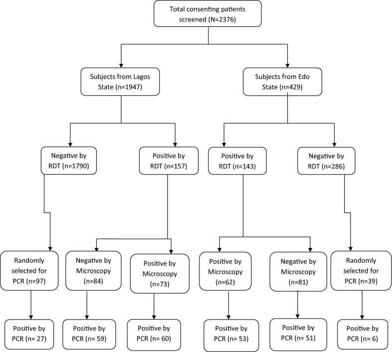
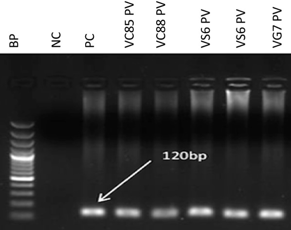
A cross-sectional survey between September 2016 and March 2017 was conducted. 436 febrile patients were included for the present work. Venous blood of these patients was subjected to RDT as well as microscopy. Further, parasite DNA was isolated from positive samples and PCR diagnostic was employed followed by direct sequencing of the 18S rRNA of Plasmodium species as well as sequencing of a portion of the promoter region of the Duffy antigen receptor for chemokines. Samples positive for P. vivax were re-amplified several times and finally using the High Fidelity Taq to rule out any bias introduced.
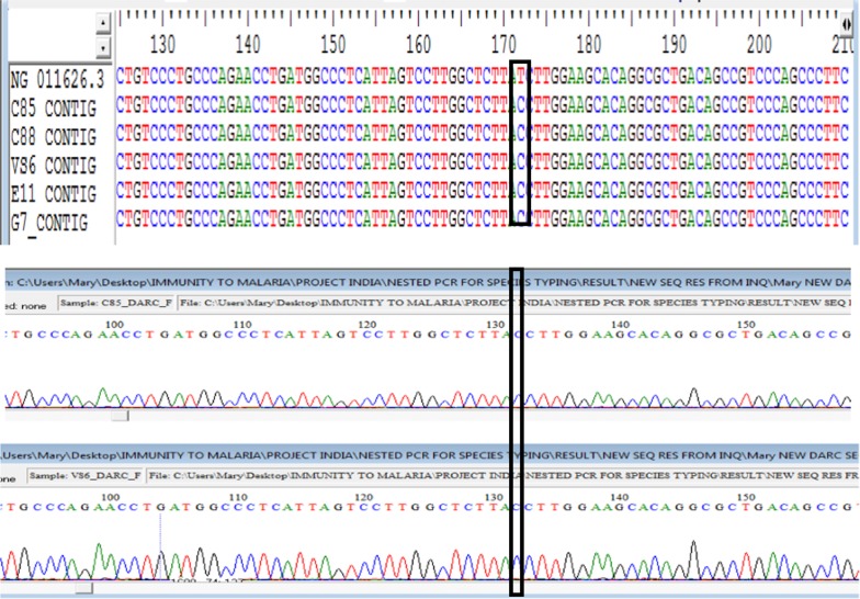
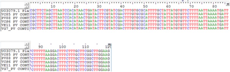
Of the 256 (58.7%) amplifiable malaria parasite DNA, P. falciparum was, as expected, the major cause of infection, either alone 85.5% (219/256; 97 from Edo and 122 from Lagos), or mixed with P. malariae 6.3% (16/256) or with P. vivax 1.6% (4/256). Only one of the five P. vivax isolates was found to be a single infection. DNA sequencing and subsequent alignment of the 18S rRNA of P. vivax with the reference strains displayed very high similarities (100%). Remarkably, the T-33C was identified in all P. vivax samples, thus confirming that all vivax-infected patients in the current study are Duffy negative.
The present study gave the first molecular evidence of P. vivax in Nigeria in Duffy negative individuals. Though restricted to two states; Edo in South-South and Lagos in South-west Nigeria, the real burden of this species in Nigeria and sub-Saharan Africa might have been underestimated, hence there is need to put in place a country-wide, as well as a sub-Saharan Africa-wide surveillance and appropriate control measures.
尼日利亚的疟疾主要由恶性疟原虫引起,其次是间日疟原虫和卵形疟原虫。由于该人群中达菲阴性基因的近乎固定,人们认为恶性疟原虫在尼日利亚特别是在撒哈拉以南非洲不存在。然而,在该次区域,经常有报道称在达菲阴性个体中存在间日疟原虫感染,包括来自与尼日利亚西部(贝宁共和国)和东部(喀麦隆)接壤的两个国家的报告。此外,还有两例来自尼日利亚土着人群的显微镜下间日疟原虫感染病例。因此,对尼日利亚西南部两个州(拉各斯州和埃多州)的循环疟原虫种类进行了分子监测。
本项横断面研究于 2016 年 9 月至 2017 年 3 月进行。共纳入 436 例发热患者。对这些患者的静脉血进行 RDT 和显微镜检查。此外,从阳性样本中分离寄生虫 DNA,采用 PCR 诊断,然后直接测序疟原虫 18S rRNA,以及趋化因子达菲抗原受体启动子区域的一部分测序。对间日疟原虫阳性样本进行多次扩增,最后使用高保真 Taq 排除任何引入的偏差。
在所检测的 256 份(58.7%)可扩增疟原虫 DNA 中,恶性疟原虫是感染的主要原因,单独感染占 85.5%(219/256;97 例来自埃多州,122 例来自拉各斯州),或与间日疟原虫混合感染占 6.3%(16/256),或与间日疟原虫混合感染占 1.6%(4/256)。5 株间日疟原虫分离株中只有 1 株为单纯感染。对间日疟原虫 18S rRNA 的 DNA 测序和随后的序列比对显示出非常高的相似度(100%)。值得注意的是,所有间日疟原虫样本均鉴定出 T-33C,这证实了本研究中所有间日疟原虫感染患者均为达菲阴性。
本研究首次在达菲阴性个体中提供了尼日利亚间日疟原虫的分子证据。尽管本研究仅限于南南的埃多州和西南的拉各斯州,但该物种在尼日利亚和撒哈拉以南非洲的实际负担可能被低估了,因此有必要在全国范围内以及撒哈拉以南非洲范围内建立监测和适当的控制措施。