Department of Cellular and Molecular Physiology, University of Liverpool, Liverpool, UK.
Centre for Preclinical Imaging, University of Liverpool, Liverpool, UK.
Stem Cell Res Ther. 2018 Nov 28;9(1):332. doi: 10.1186/s13287-018-1076-x.
Cell-based regenerative medicine therapies are now frequently tested in clinical trials. In many conditions, cell therapies are administered systemically, but there is little understanding of their fate, and adverse events are often under-reported. Currently, it is only possible to assess safety and fate of cell therapies in preclinical studies, specifically by monitoring animals longitudinally using multi-modal imaging approaches. Here, using a suite of in vivo imaging modalities to explore the fate of a range of human and murine cells, we investigate how route of administration, cell type and host immune status affect the fate of administered cells.
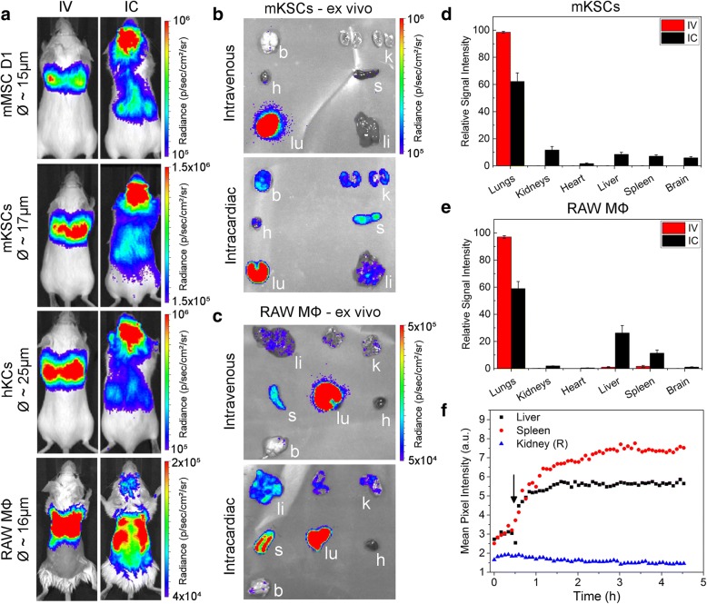
We applied a unique imaging platform combining bioluminescence, optoacoustic and magnetic resonance imaging modalities to assess the safety of different human and murine cell types by following their biodistribution and persistence in mice following administration into the venous or arterial system.
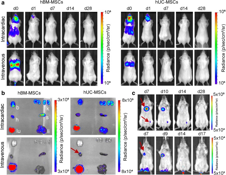
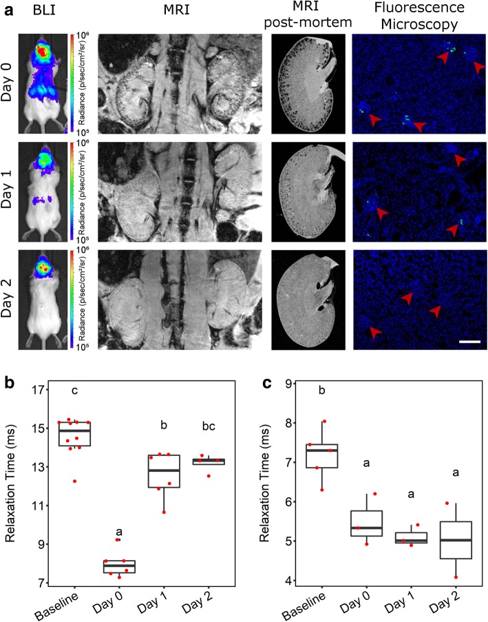
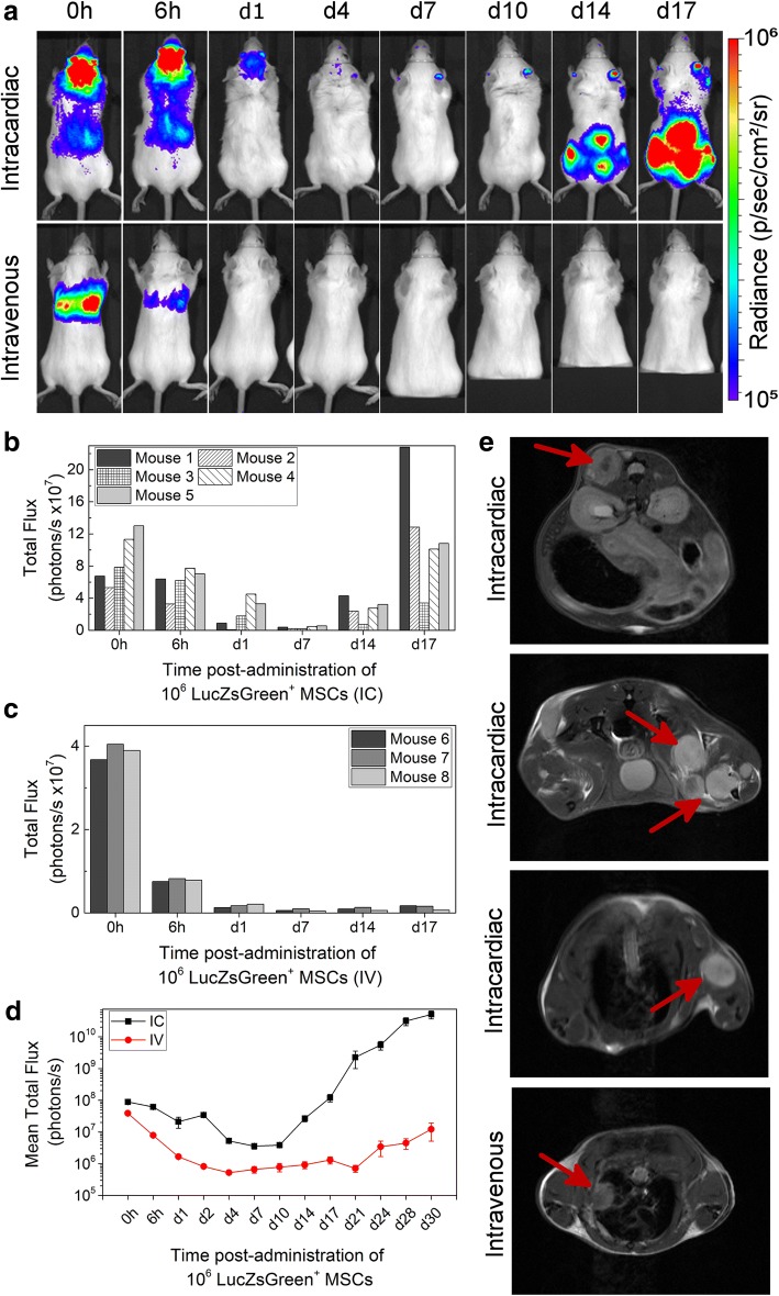
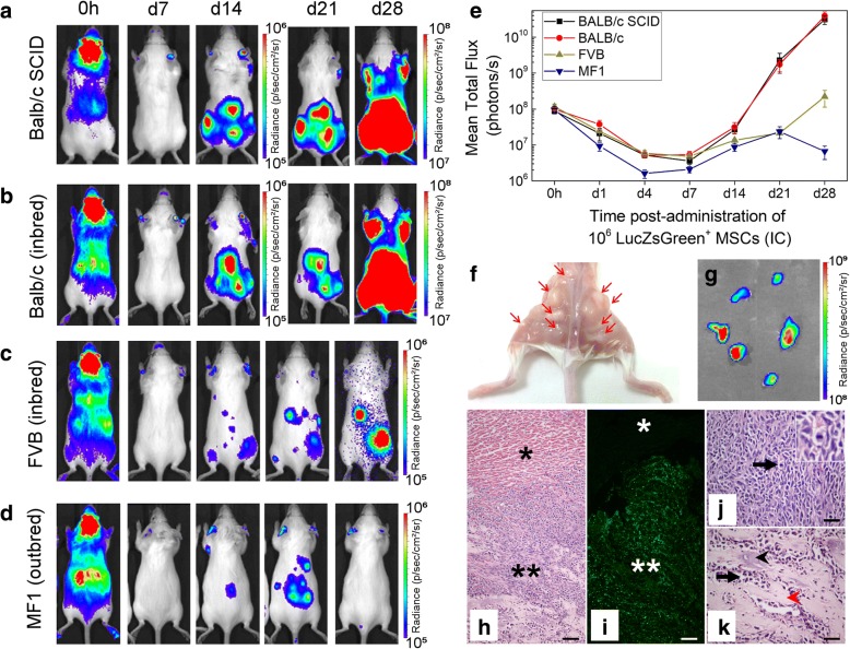
Longitudinal imaging analyses (i) suggested that the intra-arterial route may be more hazardous than intravenous administration for certain cell types, (ii) revealed that the potential of a mouse mesenchymal stem/stromal cell (MSC) line to form tumours depended on administration route and mouse strain and (iii) indicated that clinically tested human umbilical cord (hUC)-derived MSCs can transiently and unexpectedly proliferate when administered intravenously to mice.
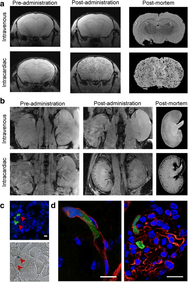
In order to perform an adequate safety assessment of potential cell-based therapies, a thorough understanding of cell biodistribution and fate post administration is required. The non-invasive imaging platform used here can expose not only the general organ distribution of these therapies, but also a detailed view of their presence within different organs and, importantly, tumourigenic potential. Our observation that the hUC-MSCs but not the human bone marrow (hBM)-derived MSCs persisted for a period in some animals suggests that therapies with these cells should proceed with caution.
基于细胞的再生医学疗法目前经常在临床试验中进行测试。在许多情况下,细胞疗法是全身性给药的,但人们对其命运知之甚少,不良事件也经常被漏报。目前,只能通过使用多模态成像方法对动物进行纵向监测,在临床前研究中评估细胞疗法的安全性和命运。在这里,我们使用一系列体内成像模式来探索一系列人类和鼠细胞的命运,研究给药途径、细胞类型和宿主免疫状态如何影响给药细胞的命运。
我们应用了一种独特的成像平台,结合生物发光、光声和磁共振成像模式,通过在静脉或动脉系统内给药后,评估不同人类和鼠细胞类型的生物分布和持久性,来评估其安全性。
纵向成像分析表明,对于某些细胞类型,动脉内途径可能比静脉内给药更危险;揭示了一种鼠间充质干细胞(MSC)系形成肿瘤的潜力取决于给药途径和鼠种;并表明,静脉内给予鼠时,经过临床测试的人脐带(hUC)衍生 MSC 可以短暂且意外地增殖。
为了对潜在的基于细胞的疗法进行充分的安全性评估,需要充分了解给药后细胞的生物分布和命运。这里使用的非侵入性成像平台不仅可以暴露这些疗法的一般器官分布,还可以详细观察它们在不同器官中的存在,并且重要的是,观察它们的致瘤潜力。我们观察到 hUC-MSC 但不是 hBM 衍生 MSC 在一些动物中持续存在一段时间,这表明这些细胞的疗法应谨慎进行。