Fleury Curado Thomaz A, Pho Huy, Dergacheva Olga, Berger Slava, Lee Rachel, Freire Carla, Asherov Aya, Sennes Luis U, Mendelowitz David, Schwartz Alan R, Polotsky Vsevolod Y
Division of Pulmonary and Critical Care Medicine, Department of Medicine, Johns Hopkins University School of Medicine, Baltimore, MD, United States.
Department of Otolaryngology, University of Sao Paulo, São Paulo, Brazil.
Front Neurol. 2018 Nov 14;9:962. doi: 10.3389/fneur.2018.00962. eCollection 2018.
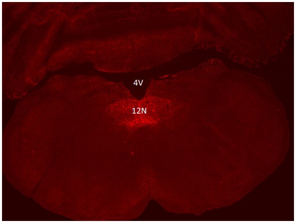
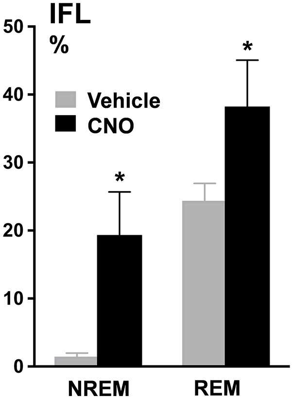
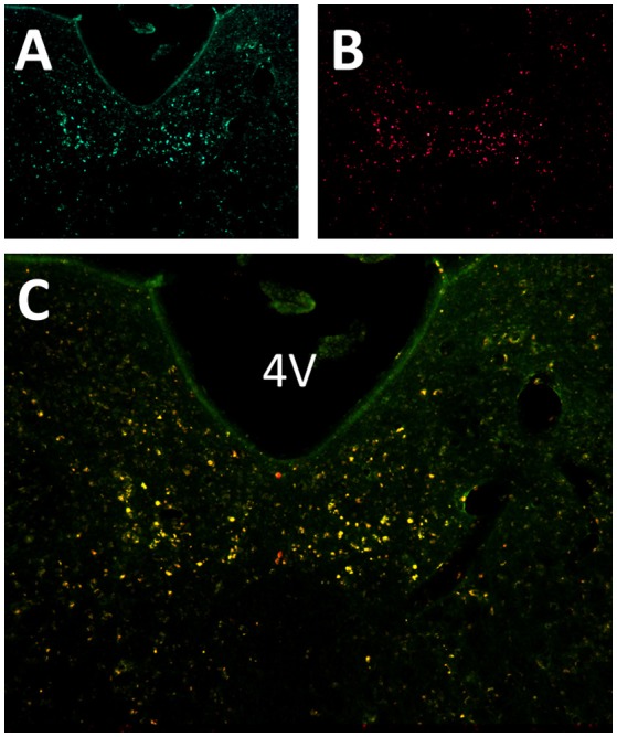
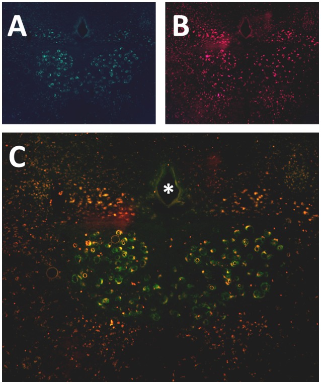
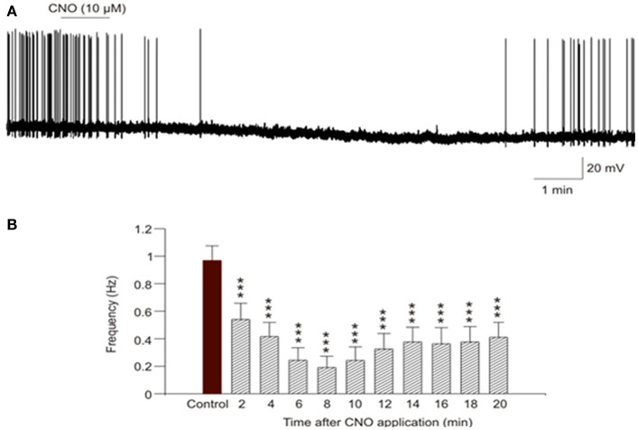
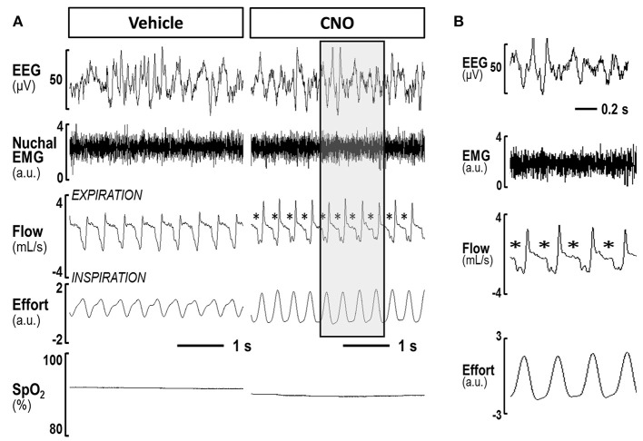
Obstructive Sleep Apnea (OSA) is a prevalent condition and a major cause of morbidity and mortality in Western Society. The loss of motor input to the tongue and specifically to the genioglossus muscle during sleep is associated with pharyngeal collapsibility and the development of OSA. We applied a novel chemogenetic method to develop a mouse model of sleep disordered breathing Our goal was to reversibly silence neuromotor input to the genioglossal muscle using an adeno-associated viral vector carrying inhibitory designer receptors exclusively activated by designer drugs AAV5-hM4Di-mCherry (DREADD), which was delivered bilaterally to the hypoglossal nucleus in fifteen C57BL/6J mice. In the experiment, 4 weeks after the viral administration mice were injected with a DREADD ligand clozapine-N-oxide (CNO, i.p., 1mg/kg) or saline followed by a sleep study; a week later treatments were alternated and a second sleep study was performed. Inspiratory flow limitation was recognized by the presence of a plateau in mid-respiratory flow; oxyhemoglobin desaturations were defined as desaturations >4% from baseline. In the electrophysiology experiment, four males and three females of 5 days of age were used. Sixteen-nineteen days after DREADD injection brain slices of medulla were prepared and individual hypoglossal motoneurons were recorded before and after CNO application. Positive mCherry staining was detected in the hypoglossal nucleus in all mice confirming successful targeting. In sleep studies, CNO markedly increased the frequency of flow limitation n NREM sleep (from 1.9 ± 1.3% after vehicle injection to 14.2 ± 3.4% after CNO, < 0.05) and REM sleep (from 22.3% ± 4.1% to 30.9 ± 4.6%, respectively, < 0.05) compared to saline treatment, but there was no significant oxyhemoglobin desaturation or sleep fragmentation. Electrophysiology recording in brain slices showed that CNO inhibited firing frequency of DREADD-containing hypoglossal motoneurons. We conclude that chemogenetic approach allows to silence hypoglossal motoneurons in mice, which leads to sleep disordered breathing manifested by inspiratory flow limitation during NREM and REM sleep without oxyhemoglobin desaturation or sleep fragmentation. Other co-morbid factors, such as compromised upper airway anatomy, may be needed to achieve recurrent pharyngeal obstruction observed in OSA.
阻塞性睡眠呼吸暂停(OSA)是一种常见病症,也是西方社会发病和死亡的主要原因。睡眠期间舌头尤其是颏舌肌运动输入的丧失与咽部可塌陷性及OSA的发展有关。我们应用一种新型化学遗传学方法建立了睡眠呼吸障碍的小鼠模型。我们的目标是使用携带仅由设计药物特异性激活的抑制性设计受体的腺相关病毒载体AAV5-hM4Di-mCherry(DREADD),可逆性地沉默颏舌肌的神经运动输入,并将其双侧注射到15只C57BL/6J小鼠的舌下神经核中。在实验中,病毒给药4周后,给小鼠注射DREADD配体氯氮平N-氧化物(CNO,腹腔注射,1mg/kg)或生理盐水,随后进行睡眠研究;一周后更换治疗方法,并进行第二次睡眠研究。吸气气流受限通过呼吸中期气流平台的出现来识别;氧合血红蛋白饱和度下降定义为相对于基线下降>4%。在电生理实验中,使用了4只5日龄雄性和3只5日龄雌性小鼠。在注射DREADD后16-19天,制备延髓脑片,并在应用CNO前后记录单个舌下运动神经元。在所有小鼠的舌下神经核中均检测到阳性mCherry染色,证实靶向成功。在睡眠研究中,与生理盐水治疗相比,CNO显著增加了非快速眼动睡眠(从注射赋形剂后的1.9±1.3%增加到注射CNO后的14.2±3.4%,P<0.05)和快速眼动睡眠(分别从22.3%±4.1%增加到3.9±4.6%,P<0.05)中气流受限的频率,但没有明显的氧合血红蛋白饱和度下降或睡眠片段化。脑片电生理记录显示,CNO抑制了含有DREADD的舌下运动神经元的放电频率。我们得出结论,化学遗传学方法可使小鼠舌下运动神经元沉默,导致非快速眼动和快速眼动睡眠期间出现以吸气气流受限为特征的睡眠呼吸障碍,而无氧合血红蛋白饱和度下降或睡眠片段化。可能需要其他共病因素,如气道解剖结构受损,才能出现OSA中观察到的复发性咽部阻塞。