Department of Radiology, Mayo Clinic and Foundation, Rochester, MN, USA; University of Oklahoma, Norman, OK, USA.
Department of Radiology, Mayo Clinic and Foundation, Rochester, MN, USA.
Neuroimage Clin. 2019;21:101605. doi: 10.1016/j.nicl.2018.11.015. Epub 2018 Nov 19.
Create an automated classifier for imaging characteristics of disproportionately enlarged subarachnoid space hydrocephalus (DESH), a neuroimaging phenotype of idiopathic normal pressure hydrocephalus (iNPH).
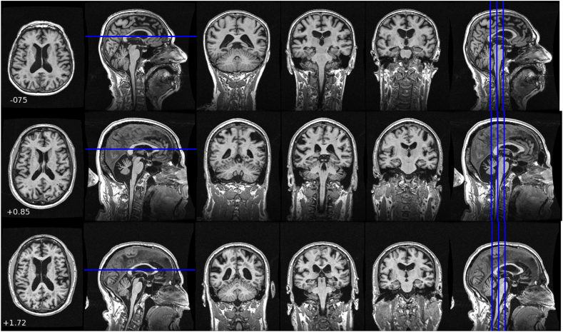
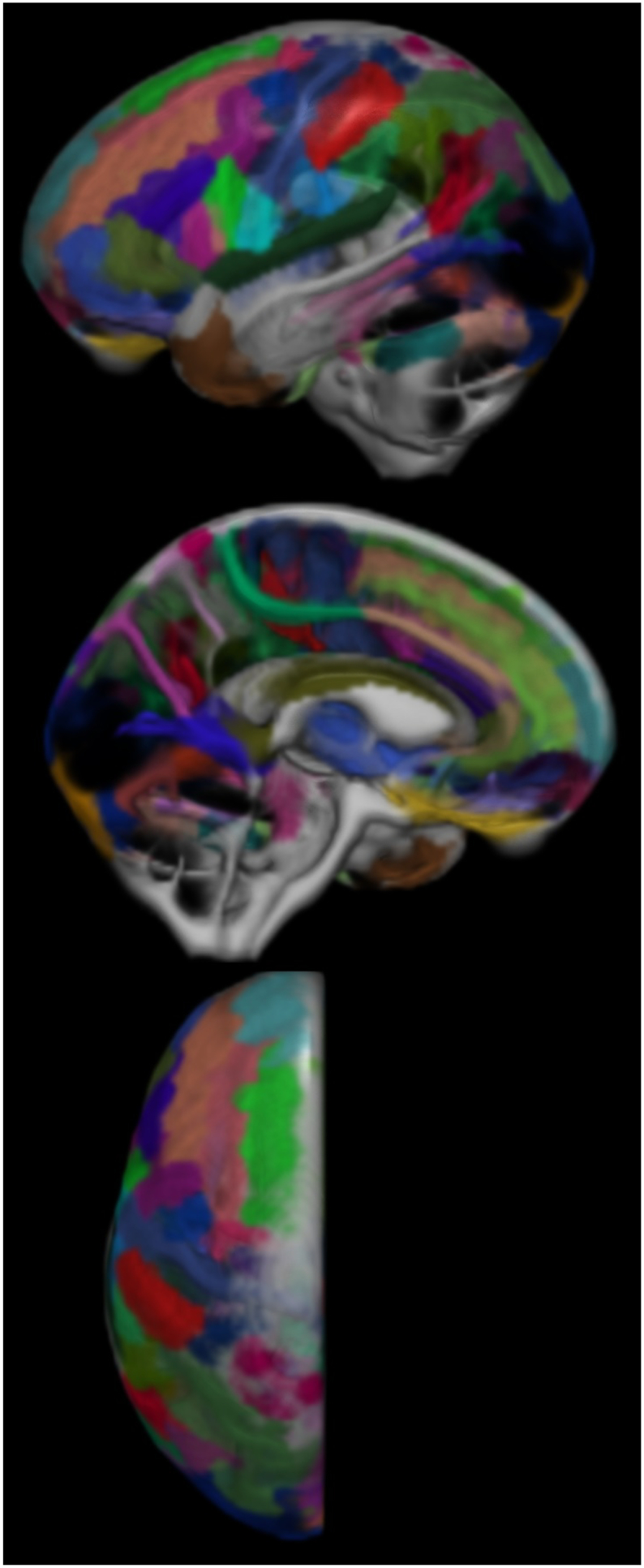
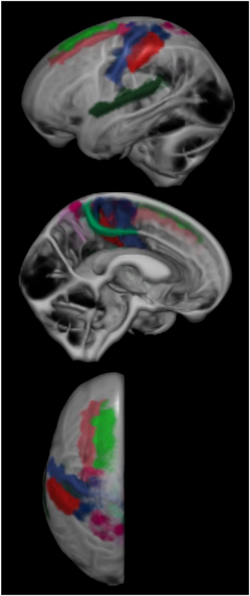
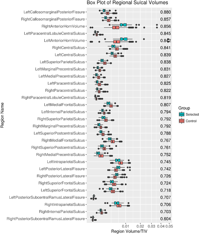
1597 patients from the Mayo Clinic Study of Aging (MCSA) were reviewed for imaging characteristics of DESH. One core feature of DESH, the presence of tightened sulci in the high-convexities (THC), was used as a surrogate for the presence of DESH as the expert clinician-defined criterion on which the classifier was trained. Anatomical MRI scans were automatically segmented for cerebrospinal fluid (CSF) and overlaid with an atlas of 123 named sulcal regions. The volume of CSF in each sulcal region was summed and normalized to total intracranial volume. Area under the receiver operating characteristic curve (AUROC) values were computed for each region individually, and these values determined feature selection for the machine learning model. Due to class imbalance in the data (72 selected scans out of 1597 total scans) adaptive synthetic sampling (a technique which generates synthetic examples based on the original data points) was used to balance the data. A support vector machine model was then trained on the regions selected.
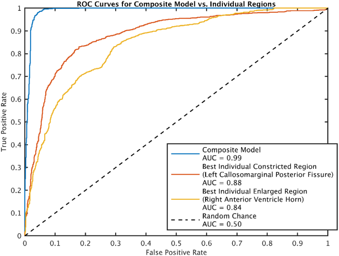
Using the automated classification model, we were able to classify scans for tightened sulci in the high convexities, as defined by the expert clinician, with an AUROC of about 0.99 (false negative ≈ 2%, false positive ≈ 5%). Ventricular volumes were among the classifier's most discriminative features but are not specific for DESH. The inclusion of regions outside the ventricles allowed specificity from atrophic neurodegenerative diseases that are also accompanied by ventricular enlargement.
Automated detection of tight high convexity, a key imaging feature of DESH, is possible by using support vector machine models with selected sulcal CSF volumes as features.
为影像学表现异常增大的蛛网膜下腔脑积水(DESH)创建一个自动分类器,DESH 是特发性正常压力脑积水(iNPH)的神经影像学表型。
对 Mayo 诊所衰老研究(MCSA)中的 1597 名患者进行 DESH 的影像学特征回顾。DESH 的一个核心特征是高凸区(THC)紧张的脑沟,将其作为 DESH 存在的替代指标,作为专家临床医生定义的分类器训练的标准。对解剖 MRI 扫描进行自动分割,以获得脑脊液(CSF)并与 123 个命名脑沟区域的图谱重叠。每个脑沟区域的 CSF 体积相加并与总颅内体积标准化。为每个区域单独计算受试者工作特征曲线(ROC)下的面积(AUROC)值,这些值确定了机器学习模型的特征选择。由于数据存在类别不平衡(1597 次总扫描中选择了 72 次扫描),因此使用自适应合成采样(一种基于原始数据点生成合成示例的技术)来平衡数据。然后在选择的区域上训练支持向量机模型。
使用自动分类模型,我们能够对专家临床医生定义的高凸区脑沟紧张进行分类,AUROC 约为 0.99(假阴性约为 2%,假阳性约为 5%)。脑室体积是分类器最具判别力的特征之一,但不是 DESH 的特异性特征。包括脑室以外的区域允许从伴有脑室扩大的萎缩性神经退行性疾病中获得特异性。
通过使用支持向量机模型并选择脑沟 CSF 体积作为特征,对 DESH 的关键影像学特征——高凸区脑沟紧张的自动检测是可行的。