Department of Biomedical and Pharmaceutical Sciences, University of Montana, 32 Campus Drive, Skaggs Building Room 284, Missoula, MT, 59812, USA.
Division of Biological Sciences, University of Montana, Missoula, MT, USA.
Arch Toxicol. 2019 Feb;93(2):355-368. doi: 10.1007/s00204-018-2366-x. Epub 2018 Nov 29.
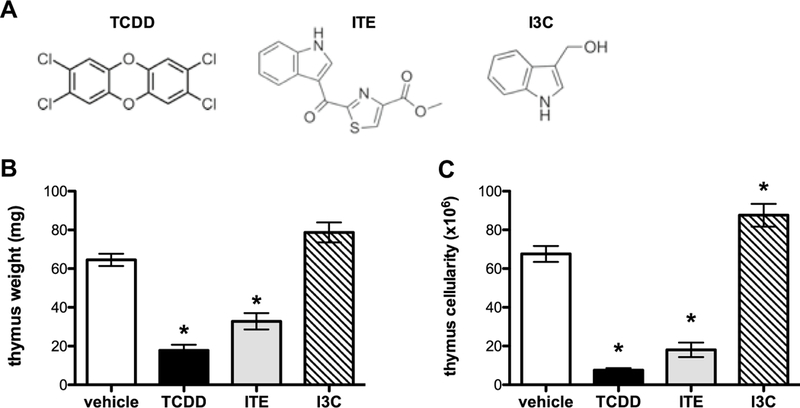
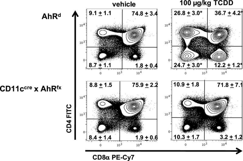
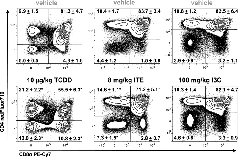
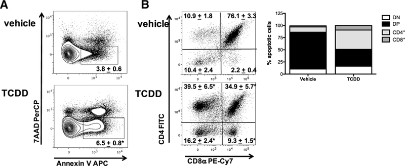
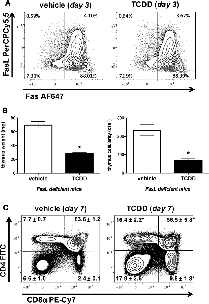
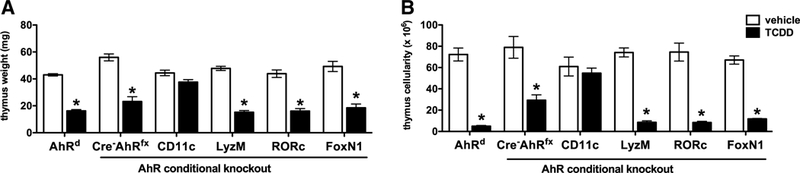
In nearly every species examined, administration of the persistent environmental pollutant, 2,3,7,8-tetrachlorodibenzo-p-dioxin (dioxin, TCDD) causes profound immune suppression and thymic atrophy in an aryl hydrocarbon receptor (AhR) dependent manner. Moreover, TCDD alters the development and differentiation of thymocytes, resulting in decreases in the relative proportion and absolute number of double positive (DP, CD4CD8) thymocytes, as well as a relative enrichment in the relative proportion and absolute number of double negative (DN, CD4CD8) and single-positive (SP) CD4CD8 and CD4CD8 thymocytes. Previous studies suggested that the target for TCDD-induced thymic atrophy resides within the hemopoietic compartment and implicated apoptosis, proliferation arrest of thymic progenitors, and emigration of DN thymocytes to the periphery as potential contributors to TCDD-induced thymic atrophy. However, the precise cellular and molecular mechanisms involved remain largely unknown. Our results show that administration of 10 µg/kg TCDD and 8 mg/kg 2-(1H-indol-3-ylcarbonyl)-4-thiazolecarboxylic acid methyl ester (ITE) induced AhR-dependent thymic atrophy in mice on day 7, whereas 100 mg/kg indole 3-carbinol (I3C) did not. Though our studies demonstrate that TCDD triggers a twofold increase in the frequency of apoptotic thymocytes, TCDD-induced thymic atrophy is not dependent on Fas-FasL interactions, and thus, enhanced apoptosis is unlikely to be a major mechanistic contributor. Finally, our results show that activation of the AhR in CD11c dendritic cells is directly responsible for TCDD-induced alterations in the development and differentiation of thymocytes, which results in thymic atrophy. Collectively, these results suggest that CD11c dendritic cells play a critical role in mediating TCDD-induced thymic atrophy and disruption of T lymphocyte development and differentiation in the thymus.
在几乎所有被检查的物种中,持久性环境污染物 2,3,7,8-四氯二苯并对二恶英(二恶英,TCDD)的给药以芳烃受体(AhR)依赖性方式引起深刻的免疫抑制和胸腺萎缩。此外,TCDD 改变了胸腺细胞的发育和分化,导致双阳性(DP,CD4CD8)胸腺细胞的相对比例和绝对数量减少,以及双阴性(DN,CD4CD8)和单阳性(SP)CD4CD8 和 CD4CD8 胸腺细胞的相对比例和绝对数量增加。先前的研究表明,TCDD 诱导的胸腺萎缩的靶标位于造血细胞群中,并暗示凋亡、胸腺祖细胞增殖停滞和 DN 胸腺细胞向外周迁移可能是 TCDD 诱导的胸腺萎缩的潜在原因。然而,确切的细胞和分子机制仍知之甚少。我们的结果表明,在第 7 天,10µg/kg TCDD 和 8mg/kg 2-(1H-吲哚-3-基羰基)-4-噻唑羧酸甲酯(ITE)给药诱导了 AhR 依赖性小鼠胸腺萎缩,而 100mg/kg 吲哚 3-甲醇(I3C)则没有。尽管我们的研究表明 TCDD 引发凋亡胸腺细胞的频率增加两倍,但 TCDD 诱导的胸腺萎缩不依赖于 Fas-FasL 相互作用,因此,增强的凋亡不太可能是主要的机制贡献者。最后,我们的结果表明,CD11c 树突状细胞中 AhR 的激活是 TCDD 诱导胸腺细胞发育和分化改变导致胸腺萎缩的直接原因。总之,这些结果表明,CD11c 树突状细胞在介导 TCDD 诱导的胸腺萎缩和破坏胸腺中 T 淋巴细胞发育和分化方面发挥着关键作用。