• 文献检索
  • 文档翻译
  • 深度研究
  • 学术资讯
  • Suppr Zotero 插件Zotero 插件
  • 邀请有礼
  • 套餐&价格
  • 历史记录
应用&插件
Suppr Zotero 插件Zotero 插件浏览器插件Mac 客户端Windows 客户端微信小程序
定价
高级版会员购买积分包购买API积分包
服务
文献检索文档翻译深度研究API 文档MCP 服务
关于我们
关于 Suppr公司介绍联系我们用户协议隐私条款
关注我们

Suppr 超能文献

核心技术专利:CN118964589B侵权必究
粤ICP备2023148730 号-1Suppr @ 2026

文献检索

告别复杂PubMed语法,用中文像聊天一样搜索,搜遍4000万医学文献。AI智能推荐,让科研检索更轻松。

立即免费搜索

文件翻译

保留排版,准确专业,支持PDF/Word/PPT等文件格式,支持 12+语言互译。

免费翻译文档

深度研究

AI帮你快速写综述,25分钟生成高质量综述,智能提取关键信息,辅助科研写作。

立即免费体验

胆小管周围腺在严重胆管损伤后人类胆管上皮再生中起关键作用。

Peribiliary Glands Are Key in Regeneration of the Human Biliary Epithelium After Severe Bile Duct Injury.

机构信息

Section of Hepatobiliary Surgery and Liver Transplantation, Department of Surgery, University of Groningen, University Medical Center Groningen, Groningen, the Netherlands.

Surgical Research Laboratory, Department of Surgery, University of Groningen, University Medical Center Groningen, Groningen, the Netherlands.

出版信息

Hepatology. 2019 Apr;69(4):1719-1734. doi: 10.1002/hep.30365. Epub 2019 Mar 5.

DOI:10.1002/hep.30365
PMID:30506902
原文链接:https://pmc.ncbi.nlm.nih.gov/articles/PMC6594148/
Abstract

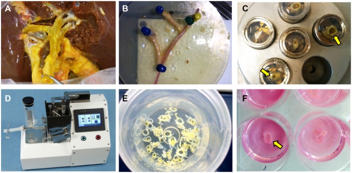
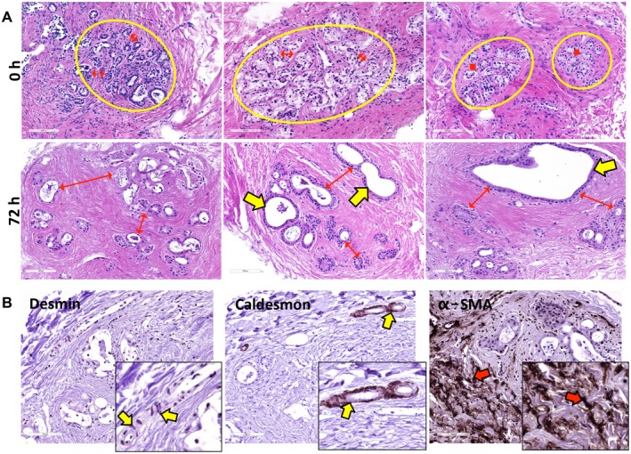
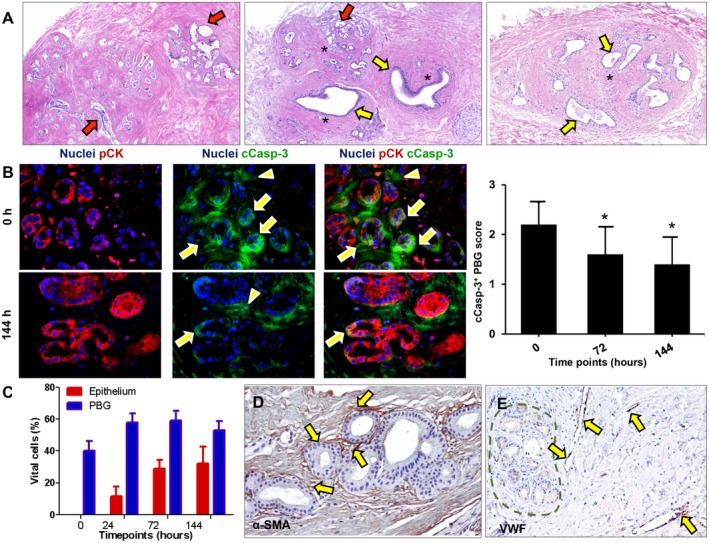
Peribiliary glands (PBG) are a source of stem/progenitor cells organized in a cellular network encircling large bile ducts. Severe cholangiopathy with loss of luminal biliary epithelium has been proposed to activate PBG, resulting in cell proliferation and differentiation to restore biliary epithelial integrity. However, formal evidence for this concept in human livers is lacking. We therefore developed an ex vivo model using precision-cut slices of extrahepatic human bile ducts obtained from discarded donor livers, providing an intact anatomical organization of cell structures, to study spatiotemporal differentiation and migration of PBG cells after severe biliary injury. Postischemic bile duct slices were incubated in oxygenated culture medium for up to a week. At baseline, severe tissue injury was evident with loss of luminal epithelial lining and mural stroma necrosis. In contrast, PBG remained relatively well preserved and different reactions of PBG were noted, including PBG dilatation, cell proliferation, and maturation. Proliferation of PBG cells increased after 24 hours of oxygenated incubation, reaching a peak after 72 hours. Proliferation of PBG cells was paralleled by a reduction in PBG apoptosis and differentiation from a primitive and pluripotent (homeobox protein Nanog+/ sex-determining region Y-box 9+) to a mature (cystic fibrosis transmembrane conductance regulator+/secretin receptor+) and activated phenotype (increased expression of hypoxia-inducible factor 1 alpha, glucose transporter 1, and vascular endothelial growth factor A). Migration of proliferating PBG cells in our ex vivo model was unorganized, but resulted in generation of epithelial monolayers at stromal surfaces. Conclusion: Human PBG contain biliary progenitor cells and are able to respond to bile duct epithelial loss with proliferation, differentiation, and maturation to restore epithelial integrity. The ex vivo spatiotemporal behavior of human PBG cells provides evidence for a pivotal role of PBG in biliary regeneration after severe injury.

摘要

胆小管周围腺(PBG)是一种以细胞网络形式存在的干细胞/祖细胞来源,围绕在大的胆管周围。人们提出,严重的胆管病变导致管腔上皮丢失会激活 PBG,引起细胞增殖和分化,从而恢复胆管上皮的完整性。然而,目前在人类肝脏中缺乏这一概念的明确证据。因此,我们建立了一种使用从废弃供体肝脏中获得的离体人外胆管的精确切割切片的体外模型,该模型提供了细胞结构的完整解剖组织,以研究严重胆管损伤后 PBG 细胞的时空分化和迁移。缺血后的胆管切片在含氧的培养介质中孵育长达一周。在基线时,明显存在严重的组织损伤,表现为管腔上皮衬里丢失和壁层基质坏死。相比之下,PBG 相对保存完好,并且观察到 PBG 的不同反应,包括 PBG 扩张、细胞增殖和成熟。在含氧孵育 24 小时后,PBG 细胞的增殖增加,在 72 小时后达到高峰。PBG 细胞的增殖伴随着 PBG 凋亡的减少和从原始多能性(同源盒蛋白 Nanog+/性别决定区 Y 框 9+)向成熟(囊性纤维化跨膜电导调节体+/分泌素受体+)和激活表型(缺氧诱导因子 1α、葡萄糖转运蛋白 1 和血管内皮生长因子 A 表达增加)的分化。在我们的体外模型中,增殖的 PBG 细胞的迁移是无组织的,但导致在基质表面生成上皮单层。结论:人 PBG 含有胆管祖细胞,并且能够对胆管上皮丢失做出反应,通过增殖、分化和成熟来恢复上皮完整性。人 PBG 细胞的体外时空行为为 PBG 在严重损伤后胆管再生中的关键作用提供了证据。

https://cdn.ncbi.nlm.nih.gov/pmc/blobs/ca33/6594148/3919862e6184/HEP-69-1719-g003.jpg
https://cdn.ncbi.nlm.nih.gov/pmc/blobs/ca33/6594148/3523b2a9ec75/HEP-69-1719-g001.jpg
https://cdn.ncbi.nlm.nih.gov/pmc/blobs/ca33/6594148/2d12ce9e7698/HEP-69-1719-g002.jpg
https://cdn.ncbi.nlm.nih.gov/pmc/blobs/ca33/6594148/3919862e6184/HEP-69-1719-g003.jpg
https://cdn.ncbi.nlm.nih.gov/pmc/blobs/ca33/6594148/3523b2a9ec75/HEP-69-1719-g001.jpg
https://cdn.ncbi.nlm.nih.gov/pmc/blobs/ca33/6594148/2d12ce9e7698/HEP-69-1719-g002.jpg
https://cdn.ncbi.nlm.nih.gov/pmc/blobs/ca33/6594148/3919862e6184/HEP-69-1719-g003.jpg

相似文献

1
Peribiliary Glands Are Key in Regeneration of the Human Biliary Epithelium After Severe Bile Duct Injury.胆小管周围腺在严重胆管损伤后人类胆管上皮再生中起关键作用。
Hepatology. 2019 Apr;69(4):1719-1734. doi: 10.1002/hep.30365. Epub 2019 Mar 5.
2
Repopulating the biliary tree from the peribiliary glands.从肝外胆管周围腺体再殖胆管树。
Biochim Biophys Acta Mol Basis Dis. 2018 Apr;1864(4 Pt B):1524-1531. doi: 10.1016/j.bbadis.2017.07.037. Epub 2017 Aug 2.
3
Injury to peribiliary glands and vascular plexus before liver transplantation predicts formation of non-anastomotic biliary strictures.肝移植前肝内胆管周围腺体和血管丛损伤可预测非吻合性胆管狭窄的形成。
J Hepatol. 2014 Jun;60(6):1172-9. doi: 10.1016/j.jhep.2014.02.010. Epub 2014 Feb 19.
4
Persistent biliary hypoxia and lack of regeneration are key mechanisms in the pathogenesis of posttransplant nonanastomotic strictures.持续性胆汁缺氧和再生缺乏是移植后非吻合口狭窄发病机制中的关键机制。
Hepatology. 2022 Apr;75(4):814-830. doi: 10.1002/hep.32166. Epub 2021 Dec 11.
5
Axin2 Peribiliary Glands in the Periampullary Region Generate Biliary Epithelial Stem Cells That Give Rise to Ampullary Carcinoma.胆胰管汇合部周围的 Axin2 胆管旁腺产生胆管上皮干细胞,进而引发壶腹癌。
Gastroenterology. 2021 May;160(6):2133-2148.e6. doi: 10.1053/j.gastro.2021.01.028. Epub 2021 Jan 16.
6
Restoration of Bile Duct Injury of Donor Livers During Ex Situ Normothermic Machine Perfusion.供体肝脏在体外常温机器灌注期间胆管损伤的修复。
Transplantation. 2023 Jun 1;107(6):e161-e172. doi: 10.1097/TP.0000000000004531. Epub 2023 May 23.
7
Peribiliary gland damage due to liver transplantation involves peribiliary vascular plexus and vascular endothelial growth factor.肝移植所致的胆管周围腺损伤涉及胆管周围血管丛和血管内皮生长因子。
Eur J Histochem. 2019 May 10;63(2):3022. doi: 10.4081/ejh.2019.3022.
8
Identification of intramural epithelial networks linked to peribiliary glands that express progenitor cell markers and proliferate after injury in mice.鉴定与胆小管周围腺体相关的黏膜上皮网络,这些网络在小鼠损伤后表达祖细胞标记物并增殖。
Hepatology. 2013 Oct;58(4):1486-1496. doi: 10.1002/hep.26485. Epub 2013 Aug 6.
9
Regeneration of human extrahepatic biliary epithelium: the peribiliary glands as progenitor cell compartment.人肝外胆管上皮细胞的再生:胆管周围腺体作为祖细胞隔室。
Liver Int. 2012 Apr;32(4):554-9. doi: 10.1111/j.1478-3231.2011.02721.x. Epub 2011 Dec 15.
10
Peribiliary Gland Niche Participates in Biliary Tree Regeneration in Mouse and in Human Primary Sclerosing Cholangitis.胆小管周围腺参与小鼠胆管树再生和人类原发性硬化性胆管炎。
Hepatology. 2020 Mar;71(3):972-989. doi: 10.1002/hep.30871. Epub 2019 Oct 10.

引用本文的文献

1
A Novel Bioabsorbable Covered Stent for Advancing Bile Duct Injury Management: A Preclinical Study in a Porcine Model (With Video).一种用于推进胆管损伤管理的新型生物可吸收覆膜支架:猪模型的临床前研究(附视频)
DEN Open. 2025 Jun 10;6(1):e70162. doi: 10.1002/deo2.70162. eCollection 2026 Apr.
2
Consensus classification of biliary complications after liver transplantation: guidelines from the BileducTx meeting.肝移植术后胆道并发症的共识分类:BileducTx会议指南
Br J Surg. 2025 Apr 30;112(5). doi: 10.1093/bjs/znae321.
3
10 degree C static storage of porcine donation after circulatory death livers improves biliary viability and mitigates ischemia-reperfusion injury.

本文引用的文献

1
Hepatic Stem/Progenitor Cell Activation Differs between Primary Sclerosing and Primary Biliary Cholangitis.原发性硬化性胆管炎和原发性胆汁性胆管炎肝干细胞/祖细胞激活存在差异。
Am J Pathol. 2018 Mar;188(3):627-639. doi: 10.1016/j.ajpath.2017.11.010. Epub 2017 Dec 15.
2
Repopulating the biliary tree from the peribiliary glands.从肝外胆管周围腺体再殖胆管树。
Biochim Biophys Acta Mol Basis Dis. 2018 Apr;1864(4 Pt B):1524-1531. doi: 10.1016/j.bbadis.2017.07.037. Epub 2017 Aug 2.
3
Evaluating the antifibrotic potency of galunisertib in a human ex vivo model of liver fibrosis.
猪心脏死亡后供肝在10摄氏度静态储存可提高胆管活力并减轻缺血再灌注损伤。
Am J Transplant. 2025 Jul;25(7):1417-1431. doi: 10.1016/j.ajt.2025.03.018. Epub 2025 Mar 20.
4
Long-term machine perfusion of human split livers: a new model for regenerative and translational research.人离体肝长期机器灌流:再生和转化研究的新模式。
Nat Commun. 2024 Nov 12;15(1):9809. doi: 10.1038/s41467-024-54024-4.
5
Extrahepatic Bile Duct Organoids as a Model to Study Ischemia/Reperfusion Injury During Liver Transplantation.肝外胆管类器官作为研究肝移植中缺血/再灌注损伤的模型。
Transpl Int. 2024 Sep 11;37:13212. doi: 10.3389/ti.2024.13212. eCollection 2024.
6
Biliary fibrosis is an important but neglected pathological feature in hepatobiliary disorders: from molecular mechanisms to clinical implications.胆汁纤维化是肝胆疾病中一个重要但被忽视的病理特征:从分子机制到临床意义。
Med Rev (2021). 2024 Jul 1;4(4):326-365. doi: 10.1515/mr-2024-0029. eCollection 2024 Aug.
7
Cholangiocyte Organoids in Liver Transplantation; a Comprehensive Review.胆管细胞类器官在肝移植中的应用:全面综述。
Transpl Int. 2024 Jul 19;37:12708. doi: 10.3389/ti.2024.12708. eCollection 2024.
8
Biliary atresia.先天性胆道闭锁。
Nat Rev Dis Primers. 2024 Jul 11;10(1):47. doi: 10.1038/s41572-024-00533-x.
9
In Utero Extrahepatic Bile Duct Damage and Repair: Implications for Biliary Atresia.子宫内肝外胆管损伤与修复:对胆道闭锁的影响。
Pediatr Dev Pathol. 2024 Jul-Aug;27(4):291-310. doi: 10.1177/10935266241247479. Epub 2024 May 19.
10
Bile proteome reveals biliary regeneration during normothermic preservation of human donor livers.胆汁蛋白质组揭示了人类供体肝脏在常温保存过程中的胆管再生。
Nat Commun. 2023 Nov 30;14(1):7880. doi: 10.1038/s41467-023-43368-y.
在人肝纤维化体外模型中评估加仑替尼的抗纤维化效力。
Br J Pharmacol. 2017 Sep;174(18):3107-3117. doi: 10.1111/bph.13945. Epub 2017 Aug 11.
4
Emerging concepts in biliary repair and fibrosis.胆道修复与纤维化的新观念
Am J Physiol Gastrointest Liver Physiol. 2017 Aug 1;313(2):G102-G116. doi: 10.1152/ajpgi.00452.2016. Epub 2017 May 19.
5
DNA Damage in Stem Cells.干细胞中的 DNA 损伤。
Mol Cell. 2017 May 4;66(3):306-319. doi: 10.1016/j.molcel.2017.04.006.
6
Biliary epithelial injury-induced regenerative response by IL-33 promotes cholangiocarcinogenesis from peribiliary glands.IL-33介导的胆管上皮损伤诱导的再生反应促进胆管周围腺发生胆管癌。
Proc Natl Acad Sci U S A. 2017 May 9;114(19):E3806-E3815. doi: 10.1073/pnas.1619416114. Epub 2017 Apr 24.
7
Renal fibrosis in precision-cut kidney slices.精密切割肾切片中的肾纤维化
Eur J Pharmacol. 2016 Nov 5;790:57-61. doi: 10.1016/j.ejphar.2016.06.057. Epub 2016 Jun 30.
8
Organoids as an in vitro model of human development and disease.类器官作为人类发育和疾病的体外模型。
Nat Cell Biol. 2016 Mar;18(3):246-54. doi: 10.1038/ncb3312.
9
"Biliary Diseases with Pancreatic Counterparts": Cross-sectional Imaging Findings.“伴有胰腺对应病变的胆系疾病”:横断影像学表现。
Radiographics. 2016 Mar-Apr;36(2):374-92. doi: 10.1148/rg.2016150071. Epub 2016 Feb 1.
10
Progenitor cell niches in the human pancreatic duct system and associated pancreatic duct glands: an anatomical and immunophenotyping study.人类胰腺导管系统及相关胰腺导管腺中的祖细胞龛:一项解剖学和免疫表型研究。
J Anat. 2016 Mar;228(3):474-86. doi: 10.1111/joa.12418. Epub 2015 Nov 27.