Department of Pathology, Johns Hopkins University, Baltimore, MD, USA.
MRC Center for Regenerative Medicine, University of Edinburgh, Edinburgh, UK.
Adv Exp Med Biol. 2018;1109:21-32. doi: 10.1007/978-3-030-02601-1_3.
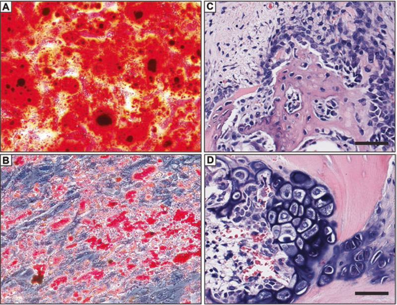
Besides seminal functions in angiogenesis and blood pressure regulation, microvascular pericytes possess a latent tissue regenerative potential that can be revealed in culture following transition into mesenchymal stem cells. Endowed with robust osteogenic potential, pericytes and other related perivascular cells extracted from adipose tissue represent a potent and abundant cell source for refined bone tissue engineering and improved cell therapies of fractures and other bone defects. The use of diverse bone formation assays in vivo, which include mouse muscle pocket osteogenesis and calvaria replenishment, rat and dog spine fusion, and rat non-union fracture healing, has confirmed the superiority of purified perivascular cells for skeletal (re)generation. As a surprising observation though, despite strong endogenous bone-forming potential, perivascular cells drive bone regeneration essentially indirectly, via recruitment by secreted factors of local osteo-progenitors.
除了在血管生成和血压调节方面的重要作用外,微血管周细胞还具有潜在的组织再生能力,在培养条件下可向间充质干细胞转分化。周细胞和从脂肪组织中提取的其他相关血管周细胞具有很强的成骨能力,是一种强大且丰富的细胞来源,可用于精细的骨组织工程和改善骨折和其他骨缺损的细胞治疗。在体内使用多种骨形成测定法,包括小鼠肌肉口袋成骨和颅骨补充、大鼠和狗脊柱融合以及大鼠骨不连愈合,证实了纯化的血管周细胞在骨骼(再)生成方面的优越性。然而,作为一个令人惊讶的观察结果,尽管具有很强的内源性成骨能力,但血管周细胞主要通过分泌因子募集局部成骨前体细胞来间接驱动骨再生。