• 文献检索
  • 文档翻译
  • 深度研究
  • 学术资讯
  • Suppr Zotero 插件Zotero 插件
  • 邀请有礼
  • 套餐&价格
  • 历史记录
应用&插件
Suppr Zotero 插件Zotero 插件浏览器插件Mac 客户端Windows 客户端微信小程序
定价
高级版会员购买积分包购买API积分包
服务
文献检索文档翻译深度研究API 文档MCP 服务
关于我们
关于 Suppr公司介绍联系我们用户协议隐私条款
关注我们

Suppr 超能文献

核心技术专利:CN118964589B侵权必究
粤ICP备2023148730 号-1Suppr @ 2026

文献检索

告别复杂PubMed语法,用中文像聊天一样搜索,搜遍4000万医学文献。AI智能推荐,让科研检索更轻松。

立即免费搜索

文件翻译

保留排版,准确专业,支持PDF/Word/PPT等文件格式,支持 12+语言互译。

免费翻译文档

深度研究

AI帮你快速写综述,25分钟生成高质量综述,智能提取关键信息,辅助科研写作。

立即免费体验

TGFβ2 诱导的腱形成作用于间充质干细胞中的钙黏蛋白和连接蛋白细胞-细胞连接蛋白。

TGFβ2-induced tenogenesis impacts cadherin and connexin cell-cell junction proteins in mesenchymal stem cells.

机构信息

Biological Engineering, 875 Perimeter Dr. MS 0904, University of Idaho, Moscow, ID, 83844, USA.

Biological Sciences, 875 Perimeter Dr. MS 3051, University of Idaho, Moscow, ID, 83844, USA.

出版信息

Biochem Biophys Res Commun. 2019 Jan 15;508(3):889-893. doi: 10.1016/j.bbrc.2018.12.023. Epub 2018 Dec 8.

DOI:10.1016/j.bbrc.2018.12.023
PMID:30538046
原文链接:https://pmc.ncbi.nlm.nih.gov/articles/PMC6394875/
Abstract

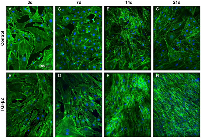
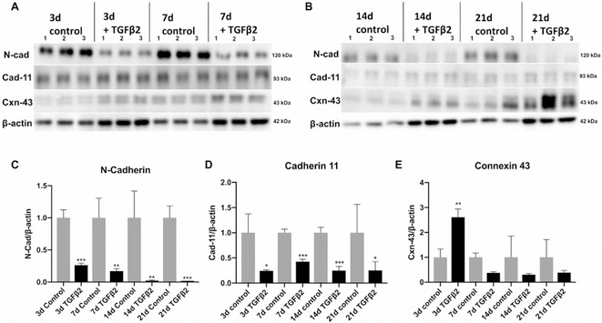
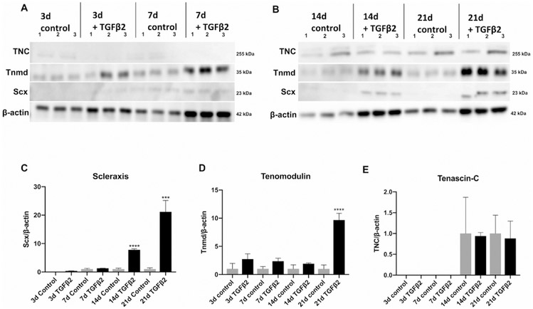
Tenogenic differentiation of stem cells is needed for tendon tissue engineering approaches. A current challenge is the limited information on the cellular-level changes during tenogenic induction. Tendon cells in embryonic and adult tendons possess an array of cell-cell junction proteins that include cadherins and connexins, but how these proteins are impacted by tenogenic differentiation is unknown. Our objective was to explore how tenogenic induction of mesenchymal stem cells (MSCs) using the transforming growth factor (TGF)β2 impacted protein markers of tendon differentiation and protein levels of N-cadherin, cadherin-11 and connexin-43. MSCs were treated with TGFβ2 for 21 days. At 3 days, TGFβ2-treated MSCs developed a fibroblastic morphology and significantly decreased levels of N-cadherin protein, which were maintained through 21 days. Similar decreases in protein levels were found for cadherin-11. Connexin-43 protein levels significantly increased at 3 days, but then decreased below control levels, though not significantly. Protein levels of scleraxis and tenomodulin were significantly increased at day 14 and 21, respectively. Taken together, our results indicate that TGFβ2 is an inducer of tendon marker proteins (scleraxis and tenomodulin) in MSCs and that tenogenesis alters the protein levels of N-cadherin, cadherin-11 and connexin-43. These findings suggest a role for connexin-43 early in tenogenesis, and show that early-onset and sustained decreases in N-cadherin and cadherin-11 may be novel markers of tenogenesis in MSCs.

摘要

干细胞的肌腱向分化对于肌腱组织工程方法是必需的。目前的挑战是肌腱向诱导过程中细胞水平变化的信息有限。胚胎和成年肌腱中的肌腱细胞具有一系列细胞-细胞连接蛋白,包括钙黏蛋白和连接蛋白,但这些蛋白如何受肌腱向分化的影响尚不清楚。我们的目的是探讨转化生长因子 (TGF)β2 对间充质干细胞 (MSCs) 的肌腱向诱导如何影响肌腱分化的蛋白标志物以及 N-钙黏蛋白、钙黏蛋白-11 和连接蛋白-43 的蛋白水平。将 MSCs 用 TGFβ2 处理 21 天。在第 3 天,TGFβ2 处理的 MSCs 表现出成纤维细胞形态,N-钙黏蛋白蛋白水平显著降低,并持续至第 21 天。钙黏蛋白-11 的蛋白水平也出现类似的降低。连接蛋白-43 的蛋白水平在第 3 天显著增加,但随后降至对照水平以下,但差异不显著。在第 14 天和第 21 天,硬骨鞘蛋白和肌腱调蛋白的蛋白水平分别显著增加。总之,我们的结果表明 TGFβ2 是 MSCs 中肌腱标志物蛋白 (硬骨鞘蛋白和肌腱调蛋白) 的诱导剂,并且肌腱发生改变了 N-钙黏蛋白、钙黏蛋白-11 和连接蛋白-43 的蛋白水平。这些发现表明连接蛋白-43 在肌腱发生早期起作用,并表明 N-钙黏蛋白和钙黏蛋白-11 的早期发生和持续减少可能是 MSCs 中肌腱发生的新标志物。

https://cdn.ncbi.nlm.nih.gov/pmc/blobs/3f48/6394875/af03b0b49e88/nihms-1516334-f0003.jpg
https://cdn.ncbi.nlm.nih.gov/pmc/blobs/3f48/6394875/6e8c8a3f59aa/nihms-1516334-f0001.jpg
https://cdn.ncbi.nlm.nih.gov/pmc/blobs/3f48/6394875/680de3e15aaf/nihms-1516334-f0002.jpg
https://cdn.ncbi.nlm.nih.gov/pmc/blobs/3f48/6394875/af03b0b49e88/nihms-1516334-f0003.jpg
https://cdn.ncbi.nlm.nih.gov/pmc/blobs/3f48/6394875/6e8c8a3f59aa/nihms-1516334-f0001.jpg
https://cdn.ncbi.nlm.nih.gov/pmc/blobs/3f48/6394875/680de3e15aaf/nihms-1516334-f0002.jpg
https://cdn.ncbi.nlm.nih.gov/pmc/blobs/3f48/6394875/af03b0b49e88/nihms-1516334-f0003.jpg

相似文献

1
TGFβ2-induced tenogenesis impacts cadherin and connexin cell-cell junction proteins in mesenchymal stem cells.TGFβ2 诱导的腱形成作用于间充质干细胞中的钙黏蛋白和连接蛋白细胞-细胞连接蛋白。
Biochem Biophys Res Commun. 2019 Jan 15;508(3):889-893. doi: 10.1016/j.bbrc.2018.12.023. Epub 2018 Dec 8.
2
Akt signaling is activated by TGFβ2 and impacts tenogenic induction of mesenchymal stem cells.Akt信号通路被转化生长因子β2(TGFβ2)激活,并影响间充质干细胞的成腱诱导。
Stem Cell Res Ther. 2021 Jan 26;12(1):88. doi: 10.1186/s13287-021-02167-2.
3
Comparative analysis of mesenchymal stem cell and embryonic tendon progenitor cell response to embryonic tendon biochemical and mechanical factors.间充质干细胞与胚胎肌腱祖细胞对胚胎肌腱生化和机械因素反应的比较分析
Stem Cell Res Ther. 2015 May 9;6(1):89. doi: 10.1186/s13287-015-0043-z.
4
Stepwise Differentiation of Mesenchymal Stem Cells Augments Tendon-Like Tissue Formation and Defect Repair In Vivo.间充质干细胞的逐步分化增强体内肌腱样组织形成和缺损修复
Stem Cells Transl Med. 2016 Aug;5(8):1106-16. doi: 10.5966/sctm.2015-0215. Epub 2016 Jun 8.
5
Mohawk promotes the tenogenesis of mesenchymal stem cells through activation of the TGFβ signaling pathway.莫霍克族通过激活转化生长因子β信号通路促进间充质干细胞的腱生成。
Stem Cells. 2015 Feb;33(2):443-55. doi: 10.1002/stem.1866.
6
Dose-Response Tendon-Specific Markers Induction by Growth Differentiation Factor-5 in Human Bone Marrow and Umbilical Cord Mesenchymal Stem Cells.生长分化因子 5 诱导人骨髓和脐带来源间充质干细胞产生肌腱特异性标志物。
Int J Mol Sci. 2020 Aug 17;21(16):5905. doi: 10.3390/ijms21165905.
7
Three-dimensional high-density co-culture with primary tenocytes induces tenogenic differentiation in mesenchymal stem cells.三维高密度共培养原代肌腱细胞可诱导间充质干细胞向肌腱细胞分化。
J Orthop Res. 2011 Sep;29(9):1351-60. doi: 10.1002/jor.21400. Epub 2011 Mar 15.
8
Role of Yes-associated protein (YAP) in regulation of mesenchymal stem cell tenogenic differentiation.Yes 相关蛋白(YAP)在间充质干细胞成肌腱分化中的调节作用。
J Mol Histol. 2022 Apr;53(2):273-283. doi: 10.1007/s10735-022-10059-9. Epub 2022 Jan 19.
9
Ex-vivo Tendon Repair Augmented with Bone Marrow Derived Mesenchymal Stem Cells Stimulated with Myostatin for Tenogenesis.用肌肉生长抑制素刺激的骨髓间充质干细胞增强体外肌腱修复以促进肌腱生成。
J Hand Surg Asian Pac Vol. 2018 Mar;23(1):47-57. doi: 10.1142/S2424835518500066.
10
Activation of AKT-mTOR Signaling Directs Tenogenesis of Mesenchymal Stem Cells.AKT-mTOR 信号的激活指导间充质干细胞的肌腱发生。
Stem Cells. 2018 Apr;36(4):527-539. doi: 10.1002/stem.2765. Epub 2018 Jan 5.

引用本文的文献

1
Biomaterial Properties and Differentiation Strategies for Tenogenic Differentiation of Mesenchymal Stem Cells.间充质干细胞腱分化的生物材料特性与分化策略
Cells. 2025 Mar 18;14(6):452. doi: 10.3390/cells14060452.
2
The LINC Complex Regulates Tendon Elastic Modulus, Collagen Crimp, and Lateral Expansion During Early Postnatal Development.LINC复合物在出生后早期发育过程中调节肌腱弹性模量、胶原卷曲和横向扩展。
J Orthop Res. 2025 Jun;43(6):1090-1100. doi: 10.1002/jor.26069. Epub 2025 Mar 16.
3
Enthesitis on Chip - A Model for Studying Acute and Chronic Inflammation of the Enthesis and its Pharmacological Treatment.

本文引用的文献

1
Mesenchymal Stem Cells: Time to Change the Name!间充质干细胞:是时候改名了!
Stem Cells Transl Med. 2017 Jun;6(6):1445-1451. doi: 10.1002/sctm.17-0051. Epub 2017 Apr 28.
2
TGFβ and FGF promote tendon progenitor fate and act downstream of muscle contraction to regulate tendon differentiation during chick limb development.转化生长因子β(TGFβ)和成纤维细胞生长因子(FGF)促进肌腱祖细胞命运,并在肌肉收缩下游发挥作用,以调节鸡胚肢体发育过程中的肌腱分化。
Development. 2016 Oct 15;143(20):3839-3851. doi: 10.1242/dev.136242. Epub 2016 Sep 13.
3
Hydrogels functionalized with N-cadherin mimetic peptide enhance osteogenesis of hMSCs by emulating the osteogenic niche.
芯片上的附着点炎——一种研究附着点急慢性炎症及其药物治疗的模型
Adv Healthc Mater. 2024 Dec;13(31):e2401815. doi: 10.1002/adhm.202401815. Epub 2024 Aug 27.
4
Recent Advances in the Use of Stem Cells in Tissue Engineering and Adjunct Therapies for Tendon Reconstruction and Future Perspectives.干细胞在组织工程中的应用进展以及肌腱重建辅助治疗和未来展望
Int J Mol Sci. 2024 Apr 19;25(8):4498. doi: 10.3390/ijms25084498.
5
Dielectrophoresis as a tool for electrophysiological characterization of stem cells.介电泳作为一种用于干细胞电生理特性表征的工具。
Biophys Rev (Melville). 2020 Dec 21;1(1):011304. doi: 10.1063/5.0025056. eCollection 2020 Dec.
6
Growth factors in the treatment of Achilles tendon injury.生长因子在跟腱损伤治疗中的应用
Front Bioeng Biotechnol. 2023 Sep 14;11:1250533. doi: 10.3389/fbioe.2023.1250533. eCollection 2023.
7
Engineering Cell-ECM-Material Interactions for Musculoskeletal Regeneration.用于肌肉骨骼再生的工程化细胞-细胞外基质-材料相互作用
Bioengineering (Basel). 2023 Apr 7;10(4):453. doi: 10.3390/bioengineering10040453.
8
Stem Cell Applications and Tenogenic Differentiation Strategies for Tendon Repair.用于肌腱修复的干细胞应用及腱分化策略
Stem Cells Int. 2023 Mar 15;2023:3656498. doi: 10.1155/2023/3656498. eCollection 2023.
9
Regulators of collagen crosslinking in developing and adult tendons.发育中和成年肌腱中胶原交联的调节剂。
Eur Cell Mater. 2022 Apr 5;43:130-152. doi: 10.22203/eCM.v043a11.
10
Axin2-lineage cells contribute to neonatal tendon regeneration.Axin2 谱系细胞有助于新生儿肌腱再生。
Connect Tissue Res. 2022 Sep;63(5):530-543. doi: 10.1080/03008207.2022.2036732. Epub 2022 Feb 18.
水凝胶通过模拟成骨龛,用 N-钙黏蛋白模拟肽功能化来增强 hMSCs 的成骨作用。
Biomaterials. 2016 Jan;77:44-52. doi: 10.1016/j.biomaterials.2015.10.072. Epub 2015 Nov 2.
4
The Role of Scleraxis in Fate Determination of Mesenchymal Stem Cells for Tenocyte Differentiation.硬骨素在间充质干细胞向肌腱细胞分化的命运决定中的作用
Sci Rep. 2015 Aug 20;5:13149. doi: 10.1038/srep13149.
5
Mechano-regulation of gap junction communications between tendon cells is dependent on the magnitude of tensile strain.肌腱细胞间缝隙连接通讯的机械调节取决于拉伸应变的大小。
Biochem Biophys Res Commun. 2015 Sep 18;465(2):281-6. doi: 10.1016/j.bbrc.2015.08.021. Epub 2015 Aug 7.
6
Comparative analysis of mesenchymal stem cell and embryonic tendon progenitor cell response to embryonic tendon biochemical and mechanical factors.间充质干细胞与胚胎肌腱祖细胞对胚胎肌腱生化和机械因素反应的比较分析
Stem Cell Res Ther. 2015 May 9;6(1):89. doi: 10.1186/s13287-015-0043-z.
7
Actin cytoskeleton contributes to the elastic modulus of embryonic tendon during early development.肌动蛋白细胞骨架在胚胎肌腱早期发育过程中对其弹性模量有贡献。
J Orthop Res. 2015 Jun;33(6):874-81. doi: 10.1002/jor.22880.
8
Cellular and molecular maturation in fetal and adult ovine calcaneal tendons.胎儿和成年绵羊跟腱的细胞与分子成熟
J Anat. 2015 Feb;226(2):126-42. doi: 10.1111/joa.12269. Epub 2014 Dec 25.
9
TGF-β1, GDF-5, and BMP-2 stimulation induces chondrogenesis in expanded human articular chondrocytes and marrow-derived stromal cells.转化生长因子-β1(TGF-β1)、生长分化因子-5(GDF-5)和骨形态发生蛋白-2(BMP-2)刺激可诱导扩增的人关节软骨细胞和骨髓来源的基质细胞发生软骨形成。
Stem Cells. 2015 Mar;33(3):762-73. doi: 10.1002/stem.1890.
10
Transcriptomic analysis of mouse limb tendon cells during development.发育过程中小鼠肢体肌腱细胞的转录组分析。
Development. 2014 Oct;141(19):3683-96. doi: 10.1242/dev.108654. Epub 2014 Sep 5.