Department of Biological Engineering, University of Idaho, 875 Perimeter Drive MS 0904, Moscow, ID, 83844-0904, USA.
School of Medicine, University of Washington, Seattle, WA, USA.
Fluids Barriers CNS. 2018 Dec 17;15(1):33. doi: 10.1186/s12987-018-0118-1.
Type 1 Chiari malformation (CM-I) has been historically defined by cerebellar tonsillar position (TP) greater than 3-5 mm below the foramen magnum (FM). Often, the radiographic findings are highly variable, which may influence the clinical course and patient outcome. In this study, we evaluate the inter-operator reliability (reproducibility) of MRI-based measurement of TP in CM-I patients and healthy controls.
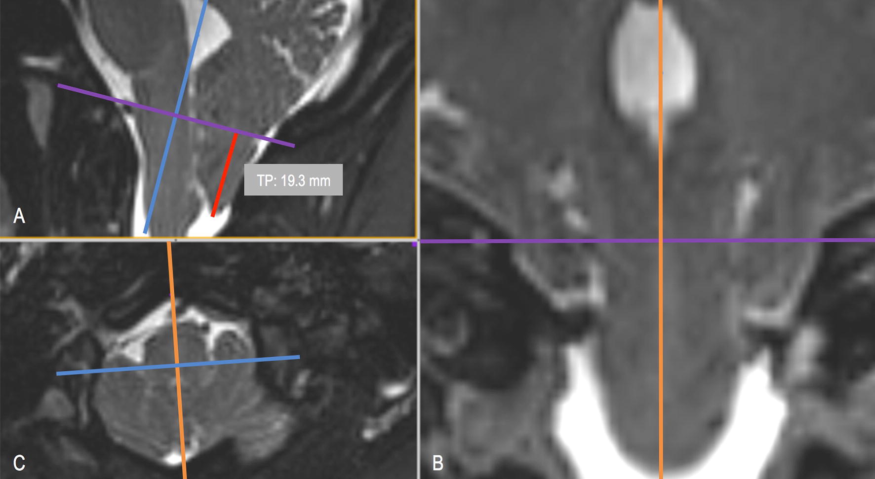
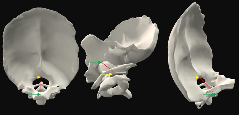
Thirty-three T2-weighted MRI sets were obtained for 23 CM-I patients (11 symptomatic and 12 asymptomatic) and 10 healthy controls. TP inferior to the FM was measured in the mid-sagittal plane by seven expert operators with reference to McRae's line. Overall agreement between the operators was quantified by intraclass correlation coefficient (ICC).
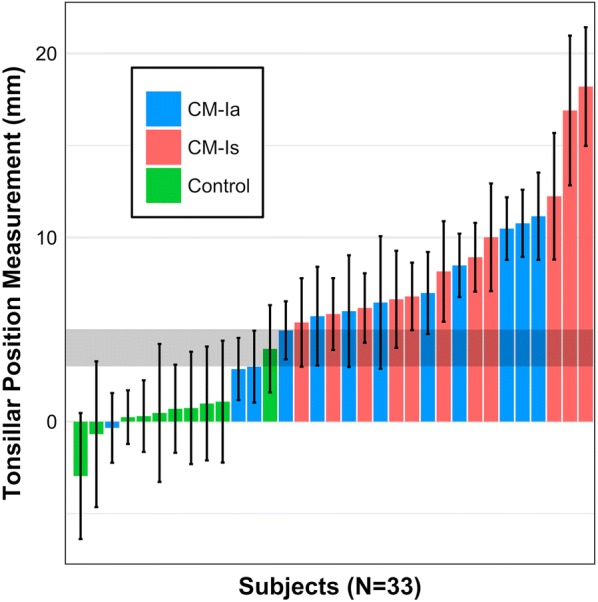
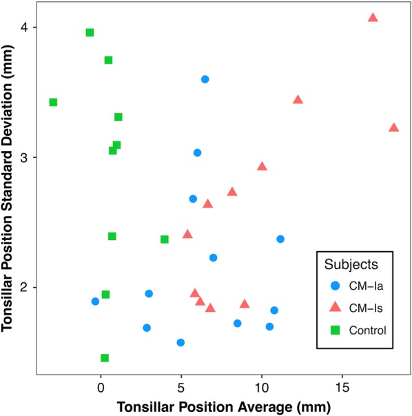
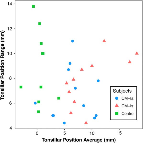
The mean and standard deviation of cerebellar TP measurements for asymptomatic (CM-Ia) and symptomatic (CM-Is) patients in mid-sagittal plane was 6.38 ± 2.19 and 9.57 ± 2.63 mm, respectively. TP measurements for healthy controls was 0.48 ± 2.88 mm. The average range of TP measurements for all data sets analyzed was 7.7 mm. Overall operator agreement for TP measurements was relatively high with an ICC of 0.83.
The results demonstrated a large average range (7.7 mm) of measurements among the seven expert operators and support that, if economically feasible, two radiologists should make independent measurements before radiologic diagnosis of CM-I and surgery is contemplated. In the future, an objective diagnostic parameter for CM-I that utilizes automated algorithms and results in smaller inter-operator variation may improve patient selection.
1 型 Chiari 畸形(CM-I)的传统定义是小脑扁桃体位置(TP)比枕骨大孔(FM)低 3-5 毫米以上。通常,放射学表现高度可变,这可能会影响临床过程和患者的预后。在这项研究中,我们评估了基于 MRI 的 CM-I 患者和健康对照组中 TP 测量的操作者间可靠性(可重复性)。
为 23 例 CM-I 患者(11 例有症状,12 例无症状)和 10 例健康对照者获得了 33 套 T2 加权 MRI。通过七位专家操作者参考 McRae 线在正中矢状面上测量小脑 TP。通过组内相关系数(ICC)来量化操作者之间的总体一致性。
无症状(CM-Ia)和有症状(CM-Is)患者在正中矢状面上小脑 TP 测量的平均值和标准差分别为 6.38±2.19 和 9.57±2.63 毫米,健康对照组为 0.48±2.88 毫米。所有分析数据集的 TP 测量平均值范围为 7.7 毫米。TP 测量的总体操作者间一致性较高,ICC 为 0.83。
结果表明,七位专家操作者的测量平均值范围较大(7.7 毫米),支持如果经济可行,在考虑 CM-I 的放射学诊断和手术之前,应由两名放射科医生进行独立测量。未来,利用自动化算法并减少操作者间差异的 CM-I 客观诊断参数可能会改善患者选择。