Zeng Jian-Jun, Wang Hai-Dong, Shen Zhong-Wei, Yao Xiao-Dong, Wu Cheng-Jun, Pan Tao
Department of Orthopaedics and Traumatology, Jiaxing Hospital of Traditional Chinese Medicine, Jiaxing, China.
Orthop Surg. 2019 Feb;11(1):117-125. doi: 10.1111/os.12412. Epub 2018 Dec 17.
To investigate the association between curcumin and the differentially expressed genes (DEG) in synovial tissues of osteoarthritis.
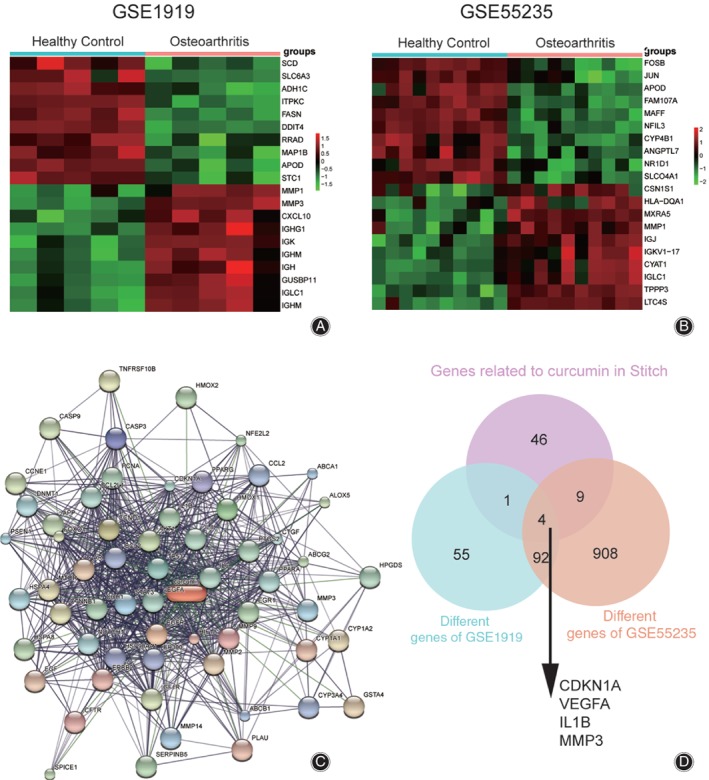
Microarray analysis was used to screen for the DEG in osteoarthritis synovial cells. Curcumin-related genes were identified through the drug-gene interaction network STITCH (http://stitch.embl.de/cgi/input.pl). Expression levels of fibronectin 1 (FN1) and collagen III protein were measured by western blot. MTT assay was used to examine the effects of different concentrations of curcumin on cell viability. Western blot and quantitative real-time polymerase chain reaction were used to validate the different expression levels of matrix metalloproteinase-3 (MMP3). Clone formation assay, flow cytometry, and the TUNEL method were conducted for detecting the cell proliferation and apoptosis rate.
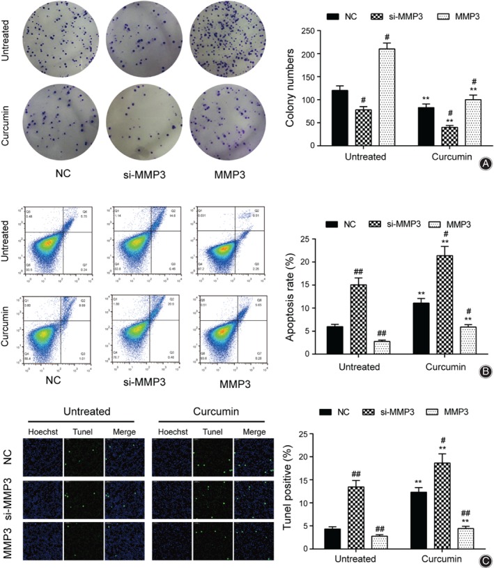
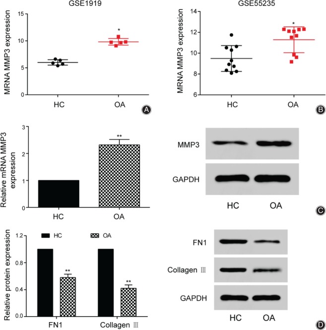
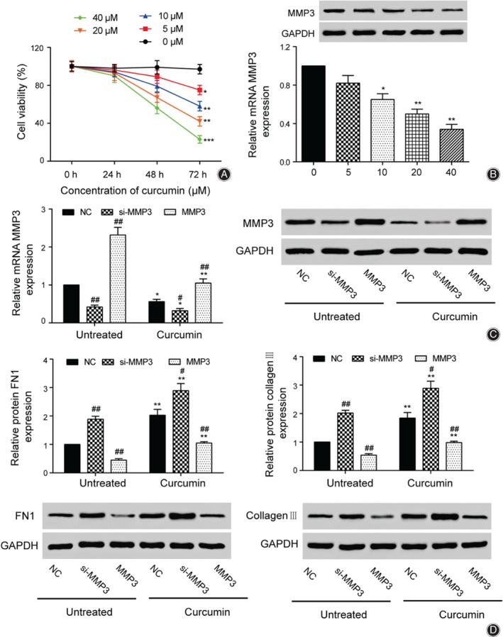
In the two chips of GSE1919 and GSE55235, the average expression of MMP3 in the osteoarthritis group was 63.7% and 12.9% higher than that of the healthy control, respectively. The results of western blot also showed that the average expression of MMP3 in 30 osteoarthritis patients was 132% higher than that of the healthy group, which confirmed that MMP3 was highly expressed in osteoarthritis group. The results of MTT showed that at 72 h, the cell viability of 40 μmol/L curcumin was the lowest and 79.6% lower than for the 0 μmol/L group, so the final curcumin concentration of 40 μmol/L was selected for subsequent experiments. Western blot results further showed that the expression of MMP3 was 44% lower in the untreated groups compared with the curcumin group, and the expressions of FN1 and collagen III were increased by 112% and 84%, respectively, which indicated that curcumin inhibited MMP3 expression and decreased osteoarthritis synovial cell activity. Cloning formation experiments showed that cell numbers increased by 75% and 20.5% in untreated and curcumin groups, and compared with the untreated group, the cells in the curcumin group decreased by 30.8%. Flow cytometry showed that the apoptotic rate in the curcumin group increased by 85.1% compared with the untreated group, but for a single group, MMP3 decreased the apoptotic rate by 53.9% and 46.7%, respectively.
MMP3 was highly expressed in osteoarthritis synovial cells. Curcumin could reduce cell viability, inhibit cell proliferation, increase cell apoptosis, and eventually alleviate inflammation of osteoarthritis by inhibiting the expression of MMP3.
探讨姜黄素与骨关节炎滑膜组织中差异表达基因(DEG)之间的关联。
采用基因芯片分析筛选骨关节炎滑膜细胞中的DEG。通过药物 - 基因相互作用网络STITCH(http://stitch.embl.de/cgi/input.pl)鉴定姜黄素相关基因。采用蛋白质免疫印迹法检测纤连蛋白1(FN1)和Ⅲ型胶原蛋白的表达水平。采用MTT法检测不同浓度姜黄素对细胞活力的影响。采用蛋白质免疫印迹法和定量实时聚合酶链反应验证基质金属蛋白酶 - 3(MMP3)的不同表达水平。进行克隆形成试验、流式细胞术和TUNEL法检测细胞增殖和凋亡率。
在GSE1919和GSE55235的两张芯片中,骨关节炎组MMP3的平均表达分别比健康对照组高63.7%和12.9%。蛋白质免疫印迹结果还显示,30例骨关节炎患者MMP3的平均表达比健康组高132%,证实MMP3在骨关节炎组中高表达。MTT结果显示,在72 h时,40 μmol/L姜黄素组的细胞活力最低,比0 μmol/L组低79.6%,因此选择40 μmol/L的最终姜黄素浓度进行后续实验。蛋白质免疫印迹结果进一步显示,与姜黄素组相比,未处理组MMP3的表达降低了44%,FN1和Ⅲ型胶原蛋白的表达分别增加了112%和84%,这表明姜黄素抑制MMP3表达并降低骨关节炎滑膜细胞活性。克隆形成实验表明,未处理组和姜黄素组的细胞数量分别增加了75%和20.5%,与未处理组相比,姜黄素组的细胞减少了30.8%。流式细胞术显示姜黄素组的凋亡率比未处理组增加了85.1%,但对于单个组,MMP3分别使凋亡率降低了53.9%和46.7%。
MMP3在骨关节炎滑膜细胞中高表达。姜黄素可降低细胞活力,抑制细胞增殖,增加细胞凋亡,并最终通过抑制MMP3的表达减轻骨关节炎炎症。