Department of Pathology and Laboratory Medicine, University of Rochester Medical Center, Rochester, NY, USA.
Konica Minolta INC., Bio Health Care Business Development Division, Corporate R&D Headquarters, No. 1 Sakura-machi, Hino-shi Tokyo, 191-8511, Japan.
BMC Cancer. 2018 Dec 18;18(1):1266. doi: 10.1186/s12885-018-5172-1.
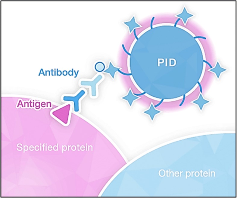
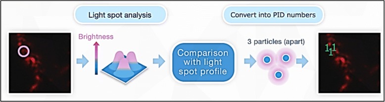
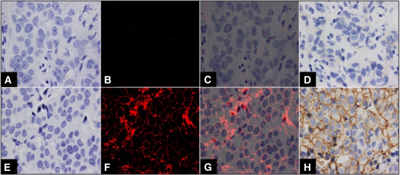
Clinical assays for the assessment of the human epidermal growth factor receptor-2 (HER2) status in breast cancer include immunohistochemistry (IHC) and in situ hybridization (ISH), both of which have limitations. Recent studies have suggested that a more quantitative approach to the measurement of HER2 protein expression may improve specificity in selecting patients for HER-2 targeted therapy. In the current study, we have used HER2 expression in breast cancer cell lines and clinical samples as a model to explore the potential utility of a novel immunodetection technique, using streptavidin coated Phosphor Integrated Dot fluorescent nanoparticles (PID), which can be quantitatively measured using computer analysis.
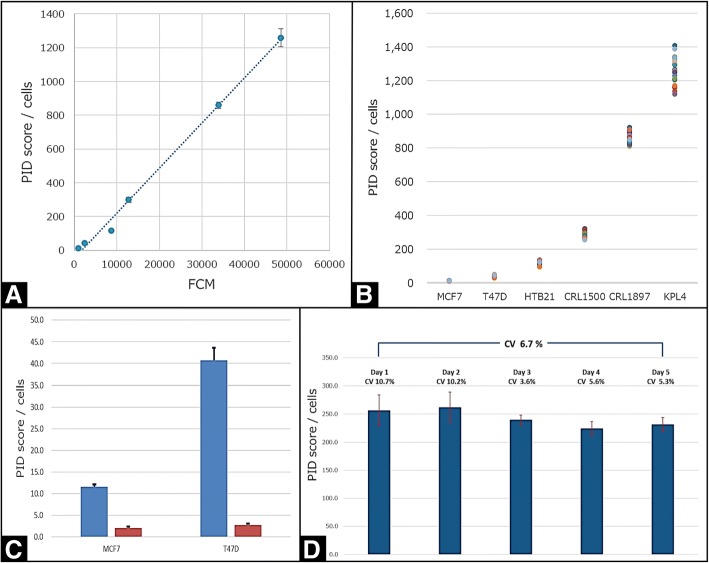
The expression of HER2 protein in cell lines was evaluated with antibody-binding capacity using fluorescence-activated cell sorting (FACS) for comparison with PID measurements to test for correlations with existing quantitative protein analysis methodologies. Various other analytic validation tests were also performed, including accuracy, precision, sensitivity, robustness and reproducibility. A methods comparison study investigated correlations between PID versus IHC and ISH in clinical samples. Lastly, we measured HER2 protein expression using PID in the pretreatment biopsies from 34 HER2-positive carcinomas that had undergone neoadjuvant trastuzumab-based chemotherapy.
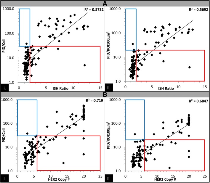
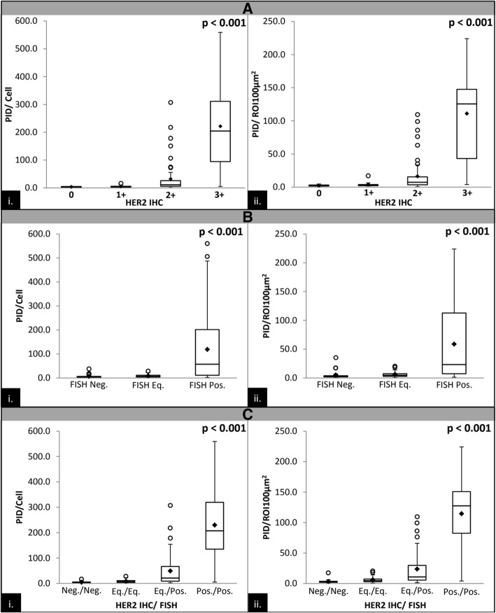
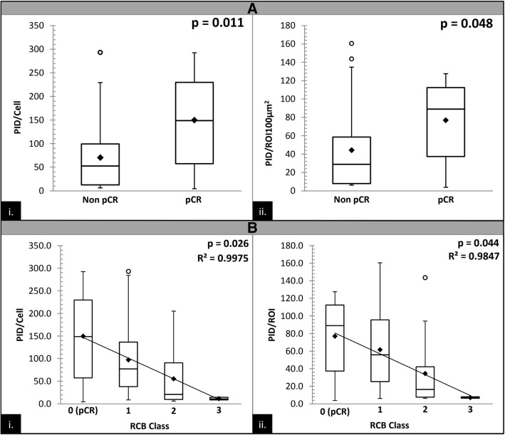
In the analytic validation, PID HER2 measurements showed a strong linear correlation with FACS analysis in breast cell lines, and demonstrated significant correlations with all aspects of precision, sensitivity, robustness and reproducibility. PID also showed strong correlations with conventional HER2 testing methodologies (IHC and ISH). In the neoadjuvant study, patients with a pathologic complete response (pCR) had a significantly higher PID score compared with patients who did not achieve a pCR (p = 0.011), and was significantly correlated to residual cancer burden (RCB) class (p = 0.026, R = 0.9975).
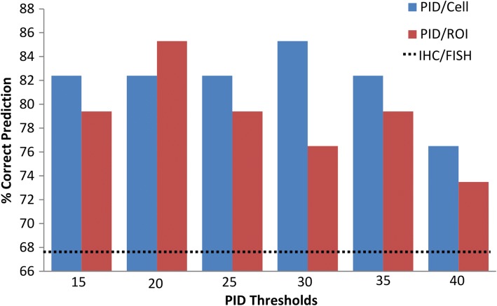
Analytic testing of PID showed that it may be a viable testing methodology that could offer advantages over other experimental or conventional biomarker diagnostic methodologies. Our data also suggests that PID quantitation of HER2 protein may offer an improvement over conventional HER2 testing in the selection of patients who will be the most likely to benefit from HER2-targeted therapy. Further studies with a larger cohort are warranted.
用于评估乳腺癌中人表皮生长因子受体 2(HER2)状态的临床检测包括免疫组织化学(IHC)和原位杂交(ISH),这两种方法都存在局限性。最近的研究表明,更定量的方法来测量 HER2 蛋白表达可能会提高选择接受 HER2 靶向治疗的患者的特异性。在本研究中,我们使用乳腺癌细胞系和临床样本中的 HER2 表达作为模型,探索一种新型免疫检测技术的潜在应用,该技术使用链霉亲和素包被的磷整合点荧光纳米颗粒(PID),可使用计算机分析进行定量测量。
使用荧光激活细胞分选(FACS)评估细胞系中 HER2 蛋白的抗体结合能力,与 PID 测量值进行比较,以测试与现有定量蛋白分析方法的相关性。还进行了各种其他分析验证测试,包括准确性、精密度、灵敏度、稳健性和重现性。方法比较研究调查了 PID 与临床样本中的 IHC 和 ISH 之间的相关性。最后,我们使用 PID 测量了 34 例接受曲妥珠单抗为基础的新辅助化疗的 HER2 阳性乳腺癌患者的预处理活检中的 HER2 蛋白表达。
在分析验证中,PID HER2 测量值与乳腺癌细胞系中的 FACS 分析显示出很强的线性相关性,并与精密度、灵敏度、稳健性和重现性的各个方面均显示出显著相关性。PID 还与传统的 HER2 检测方法(IHC 和 ISH)具有很强的相关性。在新辅助研究中,病理完全缓解(pCR)的患者的 PID 评分明显高于未达到 pCR 的患者(p=0.011),并且与残留肿瘤负荷(RCB)分级显著相关(p=0.026,R=0.9975)。
PID 的分析测试表明,它可能是一种可行的测试方法,与其他实验或传统生物标志物诊断方法相比可能具有优势。我们的数据还表明,PID 定量 HER2 蛋白可能优于传统的 HER2 检测,有助于选择最有可能从 HER2 靶向治疗中获益的患者。需要进一步的研究。