Karczmarczyk Urszula, Wojdowska Wioletta, Mikołajczak Renata, Maurin Michał, Laszuk Ewa, Garnuszek Piotr
National Centre for Nuclear Research, Radioisotope Centre POLATOM, Otwock, Poland.
Iran J Pharm Res. 2018 Fall;17(4):1201-1208.
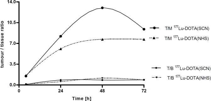
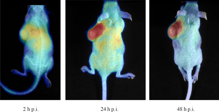
This work presents a comparative biological evaluation of Y- and Lu- labelled DOTA-SCN and DOTA-NHS conjugated to Rituximab in tumour-bearing mice. Two DOTA derivatives, p-SCN-Bn-DOTA and DOTA-NHS-ester were conjugated to Rituximab and then freeze-dried kit formulations were prepared, as previously described (1). Tissue distribution was investigated in tumour-bearing (Raji s.c.) male Rj: NMRI-Foxn1/Foxn1 mice at different time points after administration of Lu-DOTA-Rituximab or Y-DOTA-Rituximab (6 MBq/10 μg per mouse). In addition, tumour images were acquired with a PhotonIMAGER after injection of Y-DOTA (SCN)-Rituximab. All radioimmunoconjugates were obtained with high radiolabelling yield (RCP > 98%) and specific activity of ca. 0.6 GBq/mg. The conjugates were stable in human serum and in 0.9% NaCl; however, progressive aggregation was observed with time, in particular for DOTA -(SCN) conjugates. Both Lu- and Y-DOTA -(SCN)-Rituximab revealed slow blood clearance. The maximum tumour uptake was found 72 h after injection of Lu-DOTA -(SCN)-Rituximab (9.3 ID/g). A high radioactivity uptake was observed in liver and spleen, confirming the hepatobiliary excretion route. The results obtained by the radioactive optical imaging harmonize with those from the biodistribution study.
本研究对荷瘤小鼠体内与利妥昔单抗偶联的钇(Y)和镥(Lu)标记的DOTA-SCN及DOTA-NHS进行了比较生物学评价。将两种DOTA衍生物,对氨基硫脲苄基-DOTA(p-SCN-Bn-DOTA)和DOTA-NHS-酯与利妥昔单抗偶联,然后按照之前描述的方法(1)制备冻干试剂盒制剂。在给荷瘤(皮下接种Raji细胞)的雄性Rj:NMRI-Foxn1/Foxn1小鼠注射Lu-DOTA-利妥昔单抗或Y-DOTA-利妥昔单抗(每只小鼠6 MBq/10 μg)后的不同时间点,研究其组织分布。此外,注射Y-DOTA-(SCN)-利妥昔单抗后,用PhotonIMAGER采集肿瘤图像。所有放射免疫偶联物的放射性标记产率均很高(放射化学纯度>98%),比活度约为0.6 GBq/mg。这些偶联物在人血清和0.9%氯化钠溶液中稳定;然而,随着时间的推移观察到有逐渐聚集的现象,特别是对于DOTA-(SCN)偶联物。Lu-DOTA-(SCN)-利妥昔单抗和Y-DOTA-(SCN)-利妥昔单抗的血液清除均较慢。注射Lu-DOTA-(SCN)-利妥昔单抗后72小时发现肿瘤摄取量最大(9.3 ID/g)。在肝脏和脾脏中观察到高放射性摄取,证实了肝胆排泄途径。放射性光学成像获得的结果与生物分布研究的结果一致。