Lafuente Sofía, Fresno Laura, Anselmi Carlo, Lloret Albert, Espada Ivonne, Santos Laura
Foundation Hospital Veterinary Clinic, Autonomous Foundation Hospital of Barcelona, Barcelona, Spain.
Autonomous University of Barcelona, Department of Animal Medicine and Surgery, Veterinary Faculty, Barcelona, Spain.
JFMS Open Rep. 2018 Dec 16;4(2):2055116918817631. doi: 10.1177/2055116918817631. eCollection 2018 Jul-Dec.
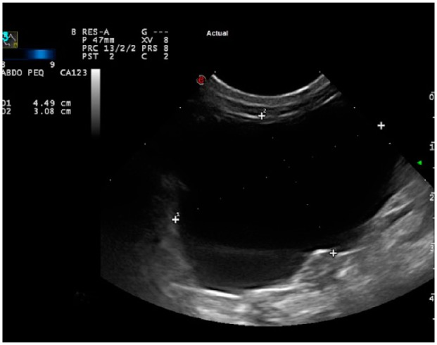
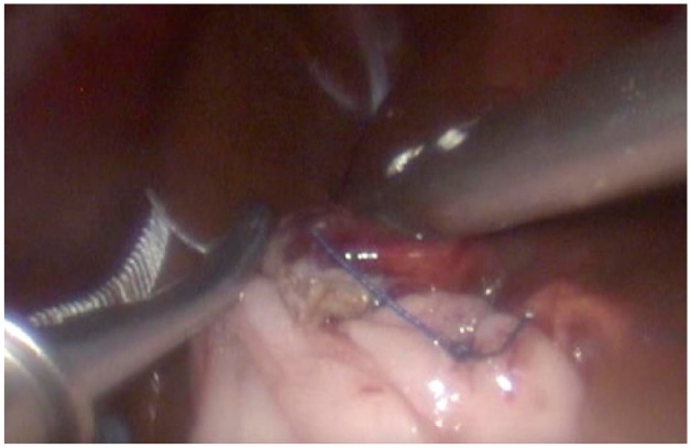
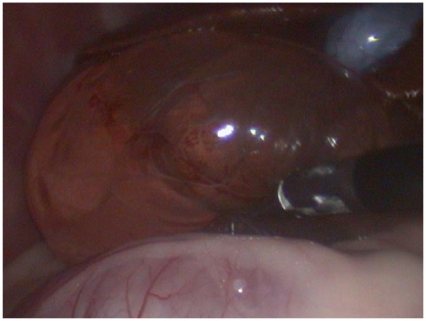
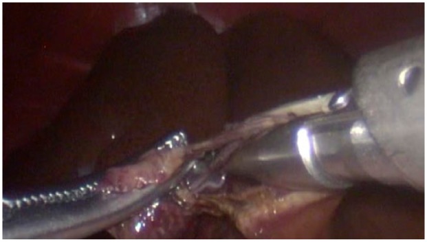
Congenital or acquired hepatic cystic lesions in cats are a rare condition. Congenital hepatic cysts are often present as part of a systemic polycystic disease involving several organs. Most cats with hepatic cysts remain clinically normal for their lives, although some patients may show abdominal distension, vomiting, abdominal pain and jaundice. An 11-year-old female neutered Persian cat was presented to our institution 3 days after the onset of inappropriate defecation and urination. This patient had a history of polycystic kidney disease and a small hepatic cystic lesion. Physical examination showed pain on abdominal palpation. Abdominal ultrasonography revealed an increase in the size of the hepatic cyst and a partial obstruction of the biliary tract. Owing to the progression of the hepatic cyst, laparoscopic excision and omentalisation were performed. The cyst was completely resected using a 5 mm laparoscopic vessel sealer/divider device. It was removed from the abdomen through one of the portals and was submitted for histological study. After cyst excision, omentopexy was performed using 4-0 USP braided absorbable material. At follow-up examination 5 days later, the physical examination was normal and abdominal palpation was not painful. A biopsy report confirmed the diagnosis of a liver cyst. A follow-up abdominal ultrasonography performed 6 months after surgery revealed no recurrence of the liver cyst.
To our knowledge, this is the first case report describing the laparoscopic technique of liver cystectomy and omentopexy in veterinary medicine. Minimally invasive surgery is gaining widespread acceptance within the veterinary community because of its benefits. However, further investigation with prospective studies are necessary.
猫的先天性或后天性肝囊性病变是一种罕见病症。先天性肝囊肿常作为涉及多个器官的全身性多囊疾病的一部分出现。大多数患有肝囊肿的猫在其一生中临床症状正常,尽管有些患者可能会出现腹胀、呕吐、腹痛和黄疸。一只11岁已绝育的雌性波斯猫在出现排便和排尿异常3天后被送至我院。该患者有多囊肾病病史及一个小的肝囊性病变。体格检查显示腹部触诊时有疼痛。腹部超声检查显示肝囊肿大小增加且胆道部分梗阻。由于肝囊肿病情进展,遂进行了腹腔镜切除及网膜固定术。使用5毫米腹腔镜血管密封/分割装置将囊肿完全切除。通过其中一个切口将其从腹部取出并送去做组织学研究。囊肿切除后,用4-0美国药典编织可吸收材料进行网膜固定术。在术后5天的随访检查中,体格检查正常且腹部触诊无疼痛。活检报告证实为肝囊肿。术后6个月进行的随访腹部超声检查显示肝囊肿未复发。
据我们所知,这是第一份描述兽医学中肝囊肿切除术及网膜固定术腹腔镜技术的病例报告。微创手术因其优点在兽医界正得到广泛认可。然而,有必要通过前瞻性研究作进一步调查。