Tanaka Aki, Hirokawa Mitsuyoshi, Higuchi Miyoko, Suzuki Ayana, Yamao Naoki, Hayashi Toshitetsu, Kuma Seiji, Miyauchi Akira
Department of Clinical Laboratory, Kuma Hospital, Kobe, Hyogo, Japan.
Department of Diagnostic Pathology and Cytology, Kuma Hospital, Kobe, Hyogo, Japan.
Diagn Cytopathol. 2019 May;47(5):452-457. doi: 10.1002/dc.24122. Epub 2018 Dec 23.
The definition of tall cell variant of papillary thyroid carcinoma (TCV-PTC) depends on the articles, and the defined cytological findings characteristic of TCV-PTC have not yet been fully analyzed. This study aimed to establish the cytological characteristics of TCV-PTC.
We retrospectively analyzed the smears of 19 TCV-PTC and 50 conventional PTC (C-PTC) cases.
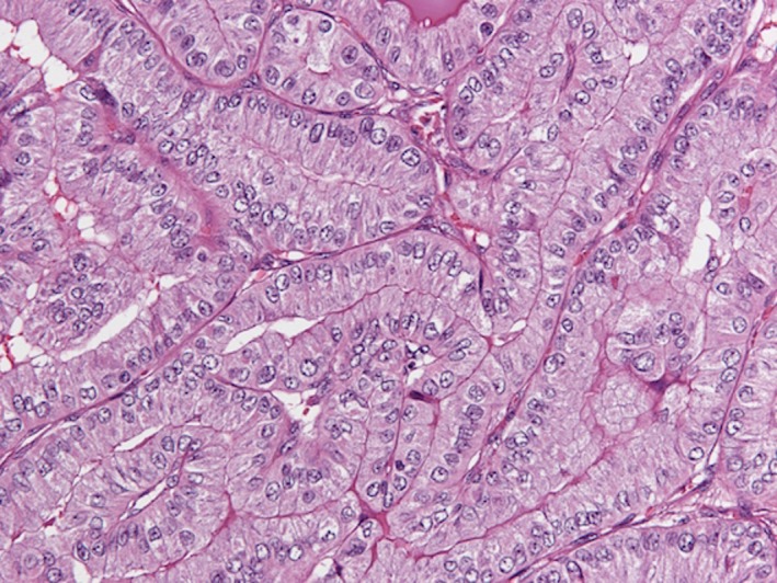
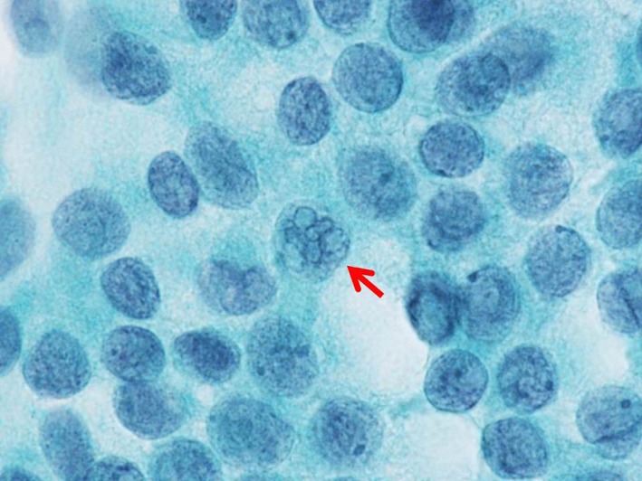
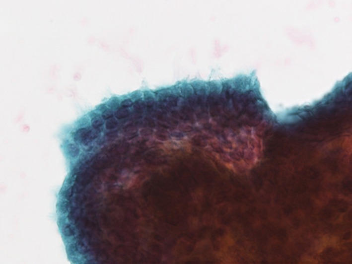
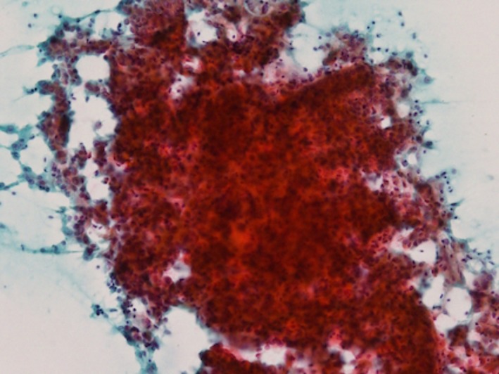
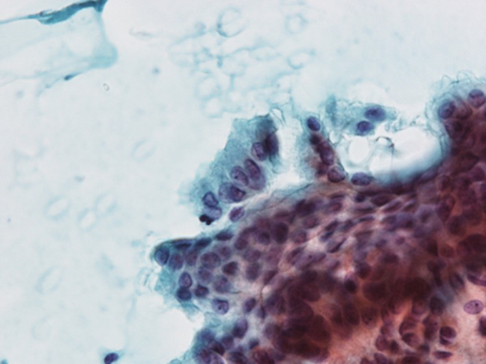
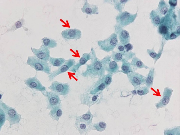
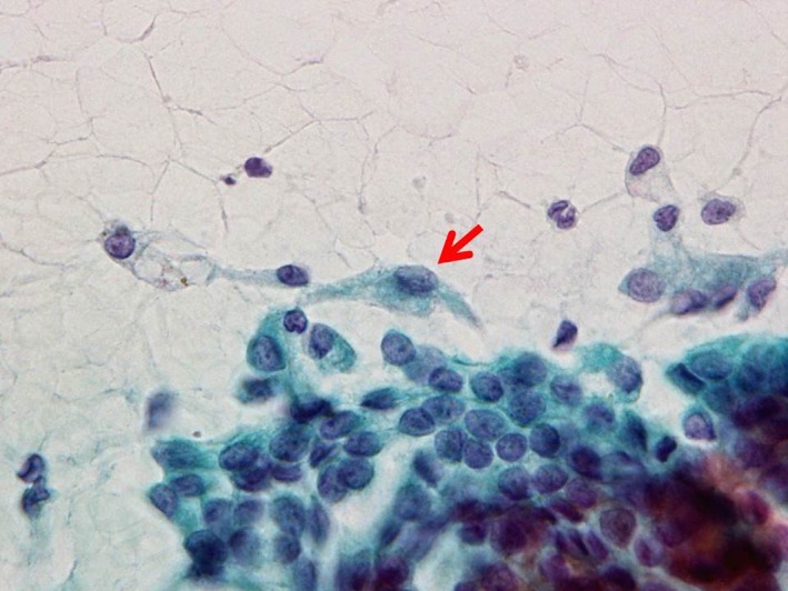
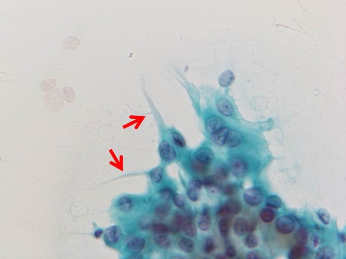
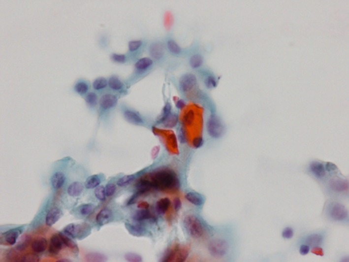
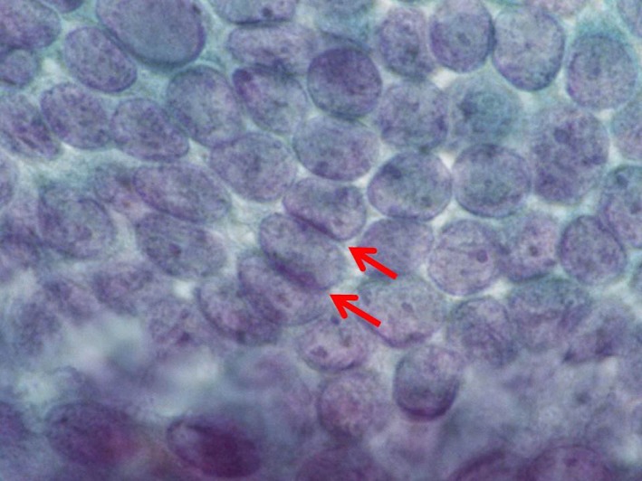
Palisaded pattern with the nuclei locating at the base of tall columnar carcinoma cells was seen in 94.7% of TCV-PTCs, and the incidence was significantly higher than that of C-PTCs (P < .0001). The palisaded pattern tended to appear at the periphery of the cell clusters. Isolated tall columnar carcinoma cells were present in 89.5% of TCV-PTCs. The incidence was significantly higher than that of C-PTC (P = .0001). Tombstone appearance was identified in 78.9% of TCV-PTCs, but not in C-PTCs. Spindle-like carcinoma cells with tapering cytoplasmic end appeared in 68.4% and 12.0% of TCV-PTC and C-PTC, respectively (P < .0001). The cytoplasm of TCV-PTC was densely stained and its cell border was distinct. Cytoplasmic elongation toward an outside of the cell clusters was observed in 89.5% of TCV-PTCs.
It is the most important to identify the presence of the tall columnar carcinoma cells on the cytological preparations, in order to distinguish TCV-PTC from C-PTC. We propose five cytological findings indicating TCV-PTC, (1) palisaded pattern, (2) tall columnar cells with the heights of at least three times their widths, (3) tombstone appearance, (4) spindle-like carcinoma cells, and (5) cytoplasmic elongation.
甲状腺乳头状癌高细胞变体(TCV-PTC)的定义因文献而异,且尚未对TCV-PTC所定义的细胞学特征进行全面分析。本研究旨在确立TCV-PTC的细胞学特征。
我们回顾性分析了19例TCV-PTC和50例传统PTC(C-PTC)病例的涂片。
94.7%的TCV-PTC可见细胞核位于高柱状癌细胞底部的栅栏状模式,其发生率显著高于C-PTC(P <.0001)。栅栏状模式倾向于出现在细胞簇的周边。89.5%的TCV-PTC存在孤立的高柱状癌细胞。其发生率显著高于C-PTC(P =.0001)。78.9%的TCV-PTC可见墓碑样外观,但C-PTC未见。分别有68.4%和12.0%的TCV-PTC和C-PTC出现带有逐渐变细的胞质末端的梭形癌细胞(P <.0001)。TCV-PTC的细胞质染色浓密,细胞边界清晰。89.5%的TCV-PTC观察到细胞质向细胞簇外部延伸。
在细胞学制片上识别高柱状癌细胞的存在对于将TCV-PTC与C-PTC区分开来最为重要。我们提出五项提示TCV-PTC的细胞学表现:(1)栅栏状模式;(2)高度至少为宽度三倍的高柱状细胞;(3)墓碑样外观;(4)梭形癌细胞;(5)细胞质延伸。