Hirokawa Mitsuyoshi, Suzuki Ayana, Miyauchi Akira
Department of Diagnostic Pathology and Cytology, Kuma Hospital, Kobe, Japan.
Department of Clinical Laboratory, Kuma Hospital, Kobe, Japan.
VideoEndocrinology. 2018 Jun 7;5(2). doi: 10.1089/ve.2018.0119. eCollection 2018.
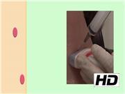
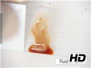
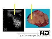
Thyroid fine-needle aspiration cytology is the most reliable preoperative diagnostic tool, but cases of failed or unsatisfactory diagnostic can occur. Therefore, we aim to improve aspiration and smearing techniques. We handle approximately 8000 thyroid fine-needle aspiration cytology cases annually. Here, we present the aspiration and smearing techniques resulting from our accumulated experience. Patients undergo aspiration cytology while seated on a barber chair, and are asked to gaze upwards to extend their anterior neck. Instead of relying on suction force, the samples are mainly obtained by cutting the tissue with needle movements. A strong negative pressure and a long aspiration time frequently produce bloody samples. Hence, we recommend negative pressure <0.3 mL and aspiration time up to 3 seconds. The obtained samples are placed on a glass slide and smeared using a second slide glass through a press and release method. When the samples are bloody, we tilt the glass slide, remove excess material, and wipe up the bloody components flowing from the slide. Liquid-based cytology is especially recommended for bloody or fluid samples. Biochemical measurement of thyroglobulin and calcitonin using fine-needle washout fluids is useful for diagnosing metastatic differentiated thyroid carcinoma and medullary thyroid carcinoma. When lymphoma is suspected, flow cytometry using aspirated samples is recommended. By applying the mentioned techniques and recommendations, we observed an increased accuracy in diagnosis and improved quality of our examinations. Fine-needle aspiration requires aspirating from the areas suitable for the diagnosis, obtaining adequate materials, and performing optimal smearing and fixation to retrieve highly accurate diagnoses. We hope our methods are helpful in improving your fine-needle aspiration cytology techniques, and result in more accurate cytological diagnoses. Thank you for taking interest in our methods. We would like to thank Dr. Louise Davies, Associate Professor of Surgery-Otolaryngology-Head and Neck Surgery, Geisel School of Medicine at Dartmouth, Hanover, NH, for her valuable comments on the creation of this video. We have no connection to any companies or products mentioned in this content. No competing financial interests exist. Runtime of video: 7 mins 15 secs The Japanese version of this video was presented at the 29th Annual Congress of the Japan Association of Endocrine Surgeons, held in May 18, 2017, in Kobe, Japan.
甲状腺细针穿刺细胞学检查是最可靠的术前诊断工具,但也可能出现诊断失败或不满意的情况。因此,我们旨在改进穿刺和涂片技术。我们每年处理约8000例甲状腺细针穿刺细胞学检查病例。在此,我们介绍基于积累经验总结出的穿刺和涂片技术。患者坐在理发椅上接受穿刺细胞学检查,并被要求向上注视以伸展其前颈部。样本主要通过针的移动切割组织来获取,而不是依靠吸力。强负压和长时间抽吸常常会产生血性样本。因此,我们建议负压<0.3 mL且抽吸时间最长为3秒。将获取的样本置于载玻片上,通过按压和松开的方法用另一张载玻片进行涂片。当样本为血性时,我们倾斜载玻片,去除多余物质,并擦去从玻片上流下来的血性成分。对于血性或液体样本,尤其推荐液基细胞学检查。使用细针冲洗液对甲状腺球蛋白和降钙素进行生化检测,有助于诊断转移性分化型甲状腺癌和甲状腺髓样癌。当怀疑为淋巴瘤时,建议对穿刺样本进行流式细胞术检测。通过应用上述技术和建议,我们观察到诊断准确性提高,检查质量得到改善。细针穿刺需要从适合诊断的区域进行抽吸,获取足够的材料,并进行最佳的涂片和固定,以获得高度准确的诊断。我们希望我们的方法有助于改进您的细针穿刺细胞学检查技术,并得出更准确的细胞学诊断结果。感谢您对我们方法的关注。我们要感谢达特茅斯盖泽尔医学院耳鼻喉头颈外科副教授路易丝·戴维斯博士对本视频制作提出的宝贵意见。我们与本内容中提及的任何公司或产品均无关联。不存在利益冲突。视频时长:7分15秒。本视频的日语版本于2017年5月18日在日本神户举行的第29届日本内分泌外科学会年会上展示。