Department of Interventional and Diagnostic Radiology, Hannover Medical School, Hannover, Germany.
Integrated Research and Treatment Center Transplantation (IfB-Tx), Hannover, Germany.
PLoS One. 2018 Dec 27;13(12):e0209103. doi: 10.1371/journal.pone.0209103. eCollection 2018.
Ex-vivo lung perfusion (EVLP) is an emerging technique promising an expansion of the donor pool and improvements in the outcome after lung transplantation. Reliable biomarkers for local assessment of organ function in the EVLP system are intensely sought after. This study aims to evaluate the feasibility of multiparametric functional magnetic resonance imaging (fMRI) in an EVLP system in a porcine aspiration model.
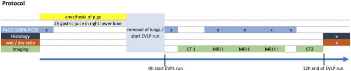
Seven female pigs were anesthetized and gastric juice was instilled in the right lower lobe bronchus to simulate aspiration. Left lungs served as control. Lungs were removed and installed in a modified EVLP system. In the 12-hour EVLP run three sequential MRI scans were performed. Oxygen-washout time, Fourier Decomposition derived ventilation and perfusion, and dynamic contrast enhanced imaging derived perfusion were calculated. PaO2:FiO2 ratio was determined and correlated. End-point histology and computed tomography served as control.
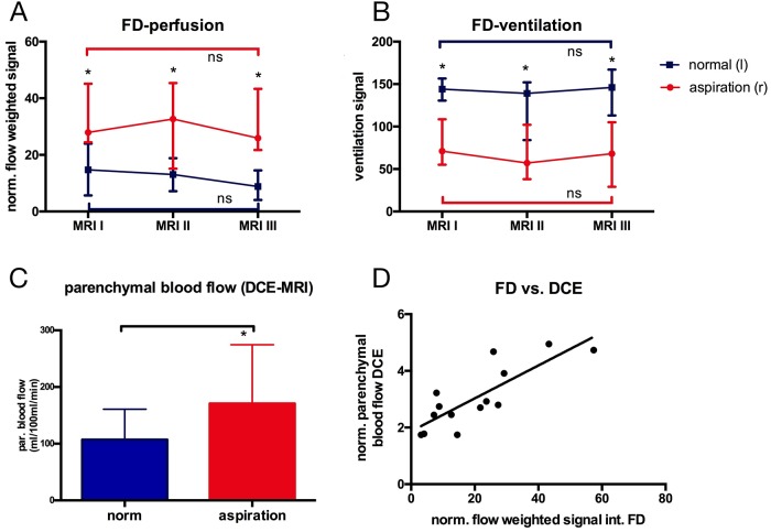
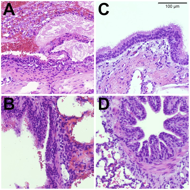
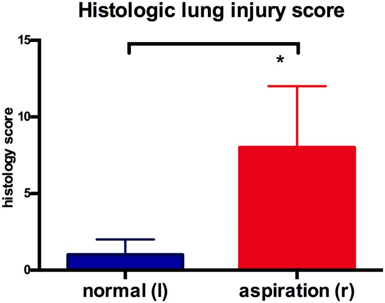
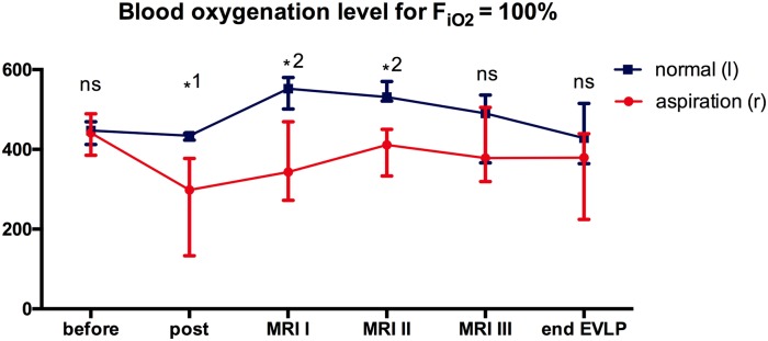
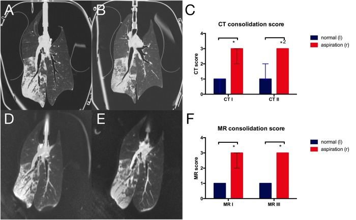
All animals completed the protocol. MRI structural images showed infiltrates in lungs after aspiration comparable to CT scans. Ventilation was significantly (p = 0.016) reduced while perfusion was increased (p = 0.016) in lungs after aspiration. Non-contrast dependent Fourier decomposition perfusion showed good correlation (R2 = 0.67) to dynamic contrast enhanced derived perfusion. Oxygen washout time was significantly increased (p = 0.016) in lungs after aspiration and showed a correlation with the PaO2:FiO2 ratio (R2 = 0.54).
Multiparametric fMRI for local assessment of organ function is feasible in EVLP and detects alterations in lung function following aspiration with correlation to clinical parameters. fMRI may improve organ assessment in ex-vivo perfusion systems, leading to a better selection of segments suitable for transplant.
体外肺灌注 (EVLP) 是一种新兴技术,有望扩大供体池并改善肺移植后的预后。人们强烈寻求用于 EVLP 系统中器官功能局部评估的可靠生物标志物。本研究旨在评估多参数功能磁共振成像 (fMRI) 在猪吸入模型 EVLP 系统中的可行性。
7 只雌性猪被麻醉,并将胃液注入右下叶支气管以模拟吸入。左肺作为对照。取出肺并安装在改良的 EVLP 系统中。在 12 小时的 EVLP 运行中,进行了三次连续的 MRI 扫描。计算了氧洗脱时间、傅里叶分解得出的通气和灌注以及动态对比增强成像得出的灌注。测定并相关了 PaO2:FiO2 比值。终点组织学和计算机断层扫描作为对照。
所有动物均完成了方案。MRI 结构图像显示吸入后肺部有浸润,与 CT 扫描相似。吸入后肺部通气明显(p = 0.016)减少,而灌注增加(p = 0.016)。非对比依赖性傅里叶分解灌注与动态对比增强衍生灌注具有良好的相关性(R2 = 0.67)。吸入后肺的氧洗脱时间明显增加(p = 0.016),与 PaO2:FiO2 比值呈相关性(R2 = 0.54)。
用于 EVLP 中器官功能局部评估的多参数 fMRI 是可行的,可检测到吸入后肺功能的改变,并与临床参数相关。fMRI 可能会改进 EVLP 系统中的器官评估,从而更好地选择适合移植的节段。