• 文献检索
  • 文档翻译
  • 深度研究
  • 学术资讯
  • Suppr Zotero 插件Zotero 插件
  • 邀请有礼
  • 套餐&价格
  • 历史记录
应用&插件
Suppr Zotero 插件Zotero 插件浏览器插件Mac 客户端Windows 客户端微信小程序
定价
高级版会员购买积分包购买API积分包
服务
文献检索文档翻译深度研究API 文档MCP 服务
关于我们
关于 Suppr公司介绍联系我们用户协议隐私条款
关注我们

Suppr 超能文献

核心技术专利:CN118964589B侵权必究
粤ICP备2023148730 号-1Suppr @ 2026

文献检索

告别复杂PubMed语法,用中文像聊天一样搜索,搜遍4000万医学文献。AI智能推荐,让科研检索更轻松。

立即免费搜索

文件翻译

保留排版,准确专业,支持PDF/Word/PPT等文件格式,支持 12+语言互译。

免费翻译文档

深度研究

AI帮你快速写综述,25分钟生成高质量综述,智能提取关键信息,辅助科研写作。

立即免费体验

一种用于动态对比增强磁共振图像分析的开源软件:重新审视UMMPerfusion

An open source software for analysis of dynamic contrast enhanced magnetic resonance images: UMMPerfusion revisited.

作者信息

Zöllner Frank G, Daab Markus, Sourbron Steven P, Schad Lothar R, Schoenberg Stefan O, Weisser Gerald

机构信息

Computer Assisted Clinical Medicine, Medical Faculty Mannheim, Heidelberg University, Theodor-Kutzer-Ufer 1-3, 68167, Mannheim, Germany.

Division of Biomedical Imaging, University of Leeds, Leeds, UK.

出版信息

BMC Med Imaging. 2016 Jan 14;16:7. doi: 10.1186/s12880-016-0109-0.

DOI:10.1186/s12880-016-0109-0
PMID:26767969
原文链接:https://pmc.ncbi.nlm.nih.gov/articles/PMC4712457/
Abstract

BACKGROUND

Perfusion imaging has become an important image based tool to derive the physiological information in various applications, like tumor diagnostics and therapy, stroke, (cardio-) vascular diseases, or functional assessment of organs. However, even after 20 years of intense research in this field, perfusion imaging still remains a research tool without a broad clinical usage. One problem is the lack of standardization in technical aspects which have to be considered for successful quantitative evaluation; the second problem is a lack of tools that allow a direct integration into the diagnostic workflow in radiology.

RESULTS

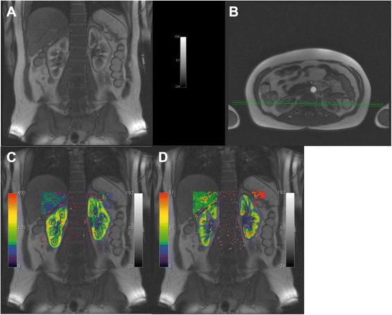
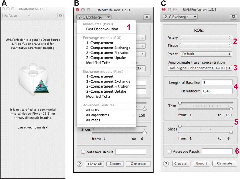
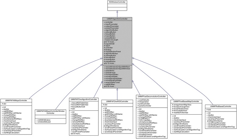
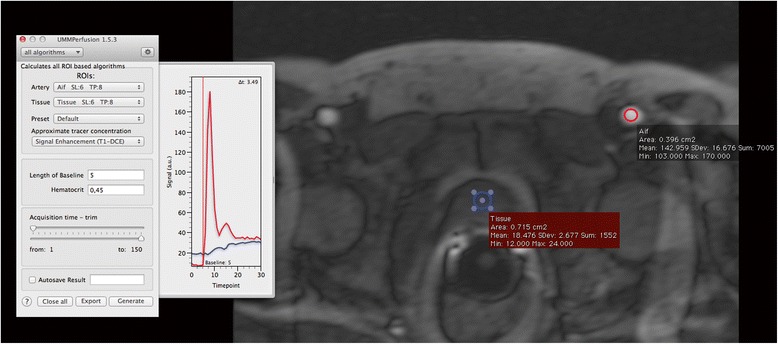
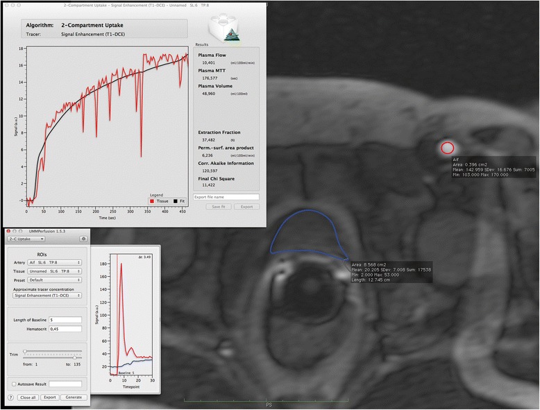
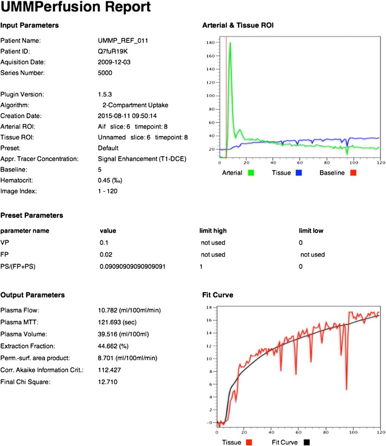
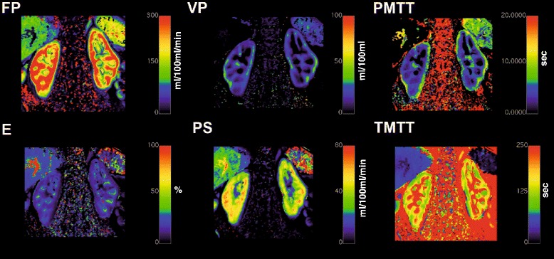
Five compartment models, namely, a one compartment model (1CP), a two compartment exchange (2CXM), a two compartment uptake model (2CUM), a two compartment filtration model (2FM) and eventually the extended Toft's model (ETM) were implemented as plugin for the DICOM workstation OsiriX. Moreover, the plugin has a clean graphical user interface and provides means for quality management during the perfusion data analysis. Based on reference test data, the implementation was validated against a reference implementation. No differences were found in the calculated parameters.

CONCLUSION

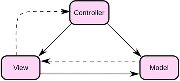
We developed open source software to analyse DCE-MRI perfusion data. The software is designed as plugin for the DICOM Workstation OsiriX. It features a clean GUI and provides a simple workflow for data analysis while it could also be seen as a toolbox providing an implementation of several recent compartment models to be applied in research tasks. Integration into the infrastructure of a radiology department is given via OsiriX. Results can be saved automatically and reports generated automatically during data analysis ensure certain quality control.

摘要

背景

灌注成像已成为一种重要的基于图像的工具,可在各种应用中获取生理信息,如肿瘤诊断与治疗、中风、(心血管)疾病或器官功能评估。然而,即使在该领域经过20年的深入研究之后,灌注成像仍然只是一种研究工具,尚未得到广泛的临床应用。一个问题是在技术方面缺乏标准化,而成功进行定量评估必须考虑这些方面;另一个问题是缺乏能够直接集成到放射诊断工作流程中的工具。

结果

五种房室模型,即单房室模型(1CP)、双房室交换模型(2CXM)、双房室摄取模型(2CUM)、双房室滤过模型(2FM)以及最终的扩展托夫特模型(ETM),被实现为DICOM工作站OsiriX的插件。此外,该插件具有简洁的图形用户界面,并在灌注数据分析过程中提供质量管理手段。基于参考测试数据,将该实现与参考实现进行了对比验证。在计算参数方面未发现差异。

结论

我们开发了用于分析DCE-MRI灌注数据的开源软件。该软件被设计为DICOM工作站OsiriX的插件。它具有简洁的图形用户界面,为数据分析提供了简单的工作流程,同时也可被视为一个工具箱,提供了几种近期房室模型的实现,可应用于研究任务。通过OsiriX可实现与放射科基础设施的集成。结果可自动保存,数据分析过程中自动生成的报告确保了一定的质量控制。

https://cdn.ncbi.nlm.nih.gov/pmc/blobs/92ea/4712457/8f38c634a62c/12880_2016_109_Fig9_HTML.jpg
https://cdn.ncbi.nlm.nih.gov/pmc/blobs/92ea/4712457/f8267311b101/12880_2016_109_Fig1_HTML.jpg
https://cdn.ncbi.nlm.nih.gov/pmc/blobs/92ea/4712457/bd45b6b64414/12880_2016_109_Fig2_HTML.jpg
https://cdn.ncbi.nlm.nih.gov/pmc/blobs/92ea/4712457/001234138f05/12880_2016_109_Fig3_HTML.jpg
https://cdn.ncbi.nlm.nih.gov/pmc/blobs/92ea/4712457/ffe3f63c61c9/12880_2016_109_Fig4_HTML.jpg
https://cdn.ncbi.nlm.nih.gov/pmc/blobs/92ea/4712457/dd2a3e519564/12880_2016_109_Fig5_HTML.jpg
https://cdn.ncbi.nlm.nih.gov/pmc/blobs/92ea/4712457/2642cf364875/12880_2016_109_Fig6_HTML.jpg
https://cdn.ncbi.nlm.nih.gov/pmc/blobs/92ea/4712457/b5935b37ae55/12880_2016_109_Fig7_HTML.jpg
https://cdn.ncbi.nlm.nih.gov/pmc/blobs/92ea/4712457/15a8650cda2d/12880_2016_109_Fig8_HTML.jpg
https://cdn.ncbi.nlm.nih.gov/pmc/blobs/92ea/4712457/8f38c634a62c/12880_2016_109_Fig9_HTML.jpg

相似文献

1
An open source software for analysis of dynamic contrast enhanced magnetic resonance images: UMMPerfusion revisited.一种用于动态对比增强磁共振图像分析的开源软件:重新审视UMMPerfusion
BMC Med Imaging. 2016 Jan 14;16:7. doi: 10.1186/s12880-016-0109-0.
2
UMMPerfusion: an open source software tool towards quantitative MRI perfusion analysis in clinical routine.UMMPerfusion:一款开源软件工具,旨在实现临床常规定量 MRI 灌注分析。
J Digit Imaging. 2013 Apr;26(2):344-52. doi: 10.1007/s10278-012-9510-6.
3
A framework for data-driven adaptive GUI generation based on DICOM.基于 DICOM 的数据驱动自适应 GUI 生成框架。
J Biomed Inform. 2018 Dec;88:37-52. doi: 10.1016/j.jbi.2018.10.009. Epub 2018 Nov 9.
4
Design and development of an ethnically-diverse imaging informatics-based eFolder system for multiple sclerosis patients.为多发性硬化症患者设计和开发基于影像信息学的多民族电子文件夹系统。
Comput Med Imaging Graph. 2015 Dec;46 Pt 2(Pt 2):257-68. doi: 10.1016/j.compmedimag.2015.09.007. Epub 2015 Oct 23.
5
Development and validation of an open source quantification tool for DSC-MRI studies.开发和验证用于 DSC-MRI 研究的开源定量工具。
Comput Biol Med. 2015 Mar;58:56-62. doi: 10.1016/j.compbiomed.2015.01.002. Epub 2015 Jan 10.
6
Contrast-agent-based perfusion MRI code repository and testing framework: ISMRM Open Science Initiative for Perfusion Imaging (OSIPI).基于对比剂的灌注 MRI 代码库和测试框架:磁共振成像学会(ISMRM)灌注成像开放科学倡议(OSIPI)。
Magn Reson Med. 2024 May;91(5):1774-1786. doi: 10.1002/mrm.29826. Epub 2023 Sep 4.
7
Advantages and Disadvantages in Image Processing with Free Software in Radiology.医学影像学中免费软件的图像处理的优缺点。
J Med Syst. 2018 Jan 15;42(3):36. doi: 10.1007/s10916-017-0888-z.
8
Validation of Interstitial Fractional Volume Quantification by Using Dynamic Contrast-Enhanced Magnetic Resonance Imaging in Porcine Skeletal Muscles.使用动态对比增强磁共振成像对猪骨骼肌间质分数体积定量的验证
Invest Radiol. 2017 Jan;52(1):66-73. doi: 10.1097/RLI.0000000000000309.
9
Dual-input two-compartment pharmacokinetic model of dynamic contrast-enhanced magnetic resonance imaging in hepatocellular carcinoma.肝细胞癌动态对比增强磁共振成像的双输入双室药代动力学模型
World J Gastroenterol. 2016 Apr 7;22(13):3652-62. doi: 10.3748/wjg.v22.i13.3652.
10
Dynamic Contrast-Enhanced Perfusion MRI of High Grade Brain Gliomas Obtained with Arterial or Venous Waveform Input Function.采用动脉或静脉波形输入函数获得的高级别脑胶质瘤动态对比增强灌注磁共振成像
J Neuroimaging. 2016 Jan-Feb;26(1):124-9. doi: 10.1111/jon.12254. Epub 2015 Apr 29.

引用本文的文献

1
A groupwise multiresolution network for DCE-MRI image registration.用于动态对比增强磁共振成像(DCE-MRI)图像配准的分组多分辨率网络。
Sci Rep. 2025 Mar 22;15(1):9891. doi: 10.1038/s41598-025-94275-9.
2
Motion-compensated image reconstruction for improved kidney function assessment using dynamic contrast-enhanced MRI.使用动态对比增强 MRI 进行运动补偿图像重建,以改善肾功能评估。
NMR Biomed. 2024 Jun;37(6):e5116. doi: 10.1002/nbm.5116. Epub 2024 Feb 15.
3
Long-term evaluation of uterine fibroid embolisation using MRI perfusion parameters and patient questionnaires: preliminary results.

本文引用的文献

1
Fitting the two-compartment model in DCE-MRI by linear inversion.通过线性反演在动态对比增强磁共振成像中拟合双室模型。
Magn Reson Med. 2016 Sep;76(3):998-1006. doi: 10.1002/mrm.25991. Epub 2015 Sep 16.
2
Dependence of DCE-MRI biomarker values on analysis algorithm.动态对比增强磁共振成像(DCE-MRI)生物标志物值对分析算法的依赖性。
PLoS One. 2015 Jul 24;10(7):e0130168. doi: 10.1371/journal.pone.0130168. eCollection 2015.
3
ROCKETSHIP: a flexible and modular software tool for the planning, processing and analysis of dynamic MRI studies.
磁共振灌注参数和患者问卷对子宫肌瘤栓塞术的长期评估:初步结果。
BMC Med Imaging. 2022 Dec 5;22(1):214. doi: 10.1186/s12880-022-00926-y.
4
Hyperpolarized [1- C]pyruvate combined with the hyperinsulinaemic euglycaemic and hypoglycaemic clamp technique in skeletal muscle in a large animal model.在大型动物模型中,将高极化 [1-13C]丙酮酸与高胰岛素正常血糖和低血糖夹技术结合应用于骨骼肌。
Exp Physiol. 2021 Dec;106(12):2412-2422. doi: 10.1113/EP089782. Epub 2021 Nov 15.
5
Analysis Protocol for Dynamic Contrast Enhanced (DCE) MRI of Renal Perfusion and Filtration.动态对比增强磁共振肾灌注和滤过分析方案。
Methods Mol Biol. 2021;2216:637-653. doi: 10.1007/978-1-0716-0978-1_38.
6
Dynamic Contrast Enhanced (DCE) MRI-Derived Renal Perfusion and Filtration: Experimental Protocol.动态对比增强(DCE)MRI 衍生的肾脏灌注和滤过:实验方案。
Methods Mol Biol. 2021;2216:429-441. doi: 10.1007/978-1-0716-0978-1_25.
7
Dynamic Contrast Enhancement (DCE) MRI-Derived Renal Perfusion and Filtration: Basic Concepts.动态对比增强磁共振成像(DCE-MRI)衍生的肾脏灌注和滤过:基本概念。
Methods Mol Biol. 2021;2216:205-227. doi: 10.1007/978-1-0716-0978-1_12.
8
MITK-ModelFit: A generic open-source framework for model fits and their exploration in medical imaging - design, implementation and application on the example of DCE-MRI.MITK-ModelFit:医学影像中模型拟合及其探索的通用开源框架 - 以 DCE-MRI 为例的设计、实现与应用
BMC Bioinformatics. 2019 Jan 16;20(1):31. doi: 10.1186/s12859-018-2588-1.
9
Multiparametric MRI for organ quality assessment in a porcine Ex-Vivo lung perfusion system.多参数 MRI 在猪离体肺灌注系统中进行器官质量评估。
PLoS One. 2018 Dec 27;13(12):e0209103. doi: 10.1371/journal.pone.0209103. eCollection 2018.
10
Variability and Reproducibility of 3-generation dual-source dynamic volume perfusion CT Parameters in Comparison to MR-perfusion Parameters in Rectal Cancer.直肠癌三代双源动态容积灌注 CT 参数与 MR 灌注参数的可变性和可重复性比较。
Sci Rep. 2018 May 2;8(1):6868. doi: 10.1038/s41598-018-25307-w.
ROCKETSHIP:一种用于动态磁共振成像研究的规划、处理和分析的灵活且模块化的软件工具。
BMC Med Imaging. 2015 Jun 16;15:19. doi: 10.1186/s12880-015-0062-3.
4
ESR Position Paper on Imaging Biobanks.红细胞沉降率(ESR)关于影像生物样本库的立场文件。
Insights Imaging. 2015 Aug;6(4):403-10. doi: 10.1007/s13244-015-0409-x. Epub 2015 May 22.
5
Comparison of the Diagnostic Performance of Four Quantitative Myocardial Perfusion Estimation Methods Used in Cardiac MR Imaging: CE-MARC Substudy.心脏磁共振成像中四种定量心肌灌注估计方法的诊断性能比较:CE-MARC子研究
Radiology. 2015 May;275(2):393-402. doi: 10.1148/radiol.14140433. Epub 2014 Dec 18.
6
Dynamic contrast-enhanced ultrasound (DCE-US) for the characterization of hepatocellular carcinoma and cholangiocellular carcinoma.动态对比增强超声(DCE-US)用于肝细胞癌和胆管细胞癌的特征描述。
Ultraschall Med. 2014 Dec;35(6):522-7. doi: 10.1055/s-0034-1385170. Epub 2014 Sep 9.
7
Pre-clinical functional Magnetic Resonance Imaging Part II: The heart.临床前功能磁共振成像 第二部分:心脏
Z Med Phys. 2014 Dec;24(4):307-22. doi: 10.1016/j.zemedi.2014.06.008. Epub 2014 Jul 9.
8
Pre-clinical functional Magnetic Resonance Imaging Part I: The kidney.临床前功能磁共振成像 第一部分:肾脏
Z Med Phys. 2014 Dec;24(4):286-306. doi: 10.1016/j.zemedi.2014.05.002. Epub 2014 Jun 12.
9
Segmentation-driven image registration- application to 4D DCE-MRI recordings of the moving kidneys.基于分割的图像配准-在移动肾脏的 4D DCE-MRI 记录中的应用。
IEEE Trans Image Process. 2014 May;23(5):2392-404. doi: 10.1109/TIP.2014.2315155.
10
Functional imaging of acute kidney injury at 3 Tesla: investigating multiple parameters using DCE-MRI and a two-compartment filtration model.3特斯拉下急性肾损伤的功能成像:使用动态对比增强磁共振成像和双室滤过模型研究多个参数
Z Med Phys. 2015 Mar;25(1):58-65. doi: 10.1016/j.zemedi.2014.01.002. Epub 2014 Mar 12.