Xiao Wei-Yuan, Zong Zhen, Qiu Man-Le, Chen Xiu-Yuan, Shen Hong-Xing, Lao Li-Feng
Department of Spine Surgery, Renji Hospital, School of Medicine, Shanghai Jiao Tong University, Shanghai, China.
Orthop Surg. 2019 Feb;11(1):126-134. doi: 10.1111/os.12414. Epub 2018 Dec 27.
To evaluate the antitumor capability and to investigate the underlying molecular mechanism of paclitaxel.
First, cck-8 and apoptosis assays were used to determine survival and apoptotic effects of HS 737.T cells under treatment of paclitaxel. Next, RNA-seq and bioinformatics were used to determine the differentially expressed genes and to analyze the pathway involved. Quantitative real-time polymerase chain reaction was used to verify the accuracy of some differentially expressed genes (DEG). ClueGO was used to decode and visualize functionally grouped GO terms of differentially expressed genes, and to map the DEG protein-protein interactions (PPI) network. Western blotting was used to check the expression of target genes, the cleavage of Caspase-3 and PARP1, and the phosphorylation level of p53. Finally, transcriptomics, bioinformatics, and RNAi were used to estimate the antitumor capability and to identify the underlying mechanisms of paclitaxel in GCTB.
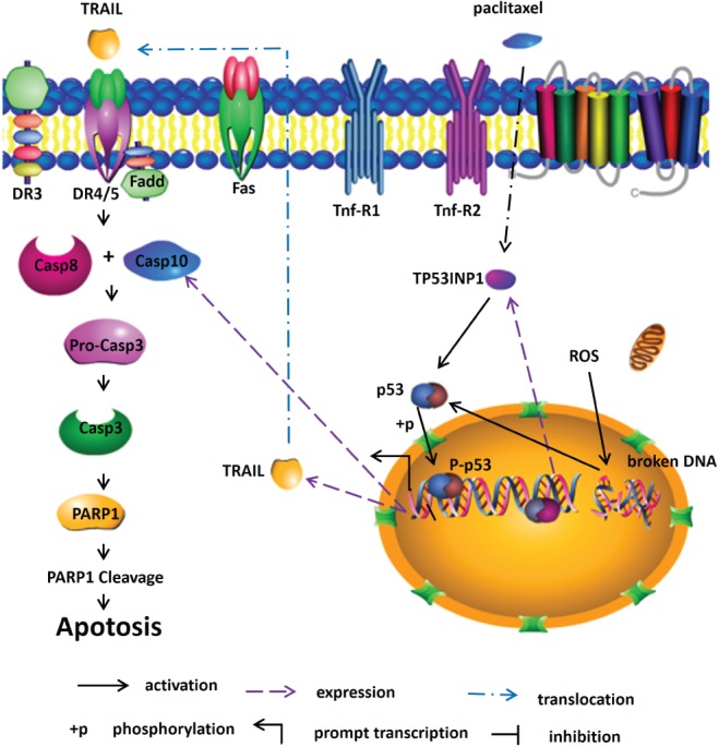
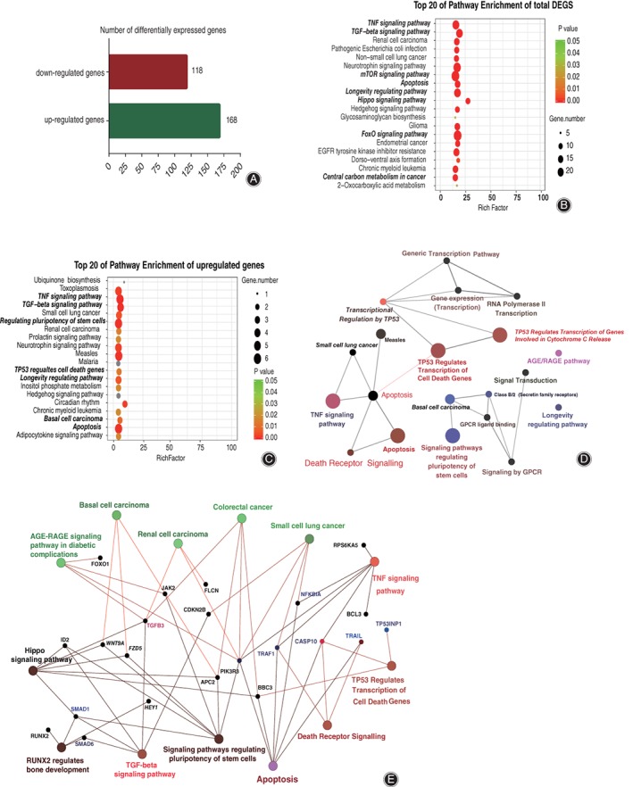
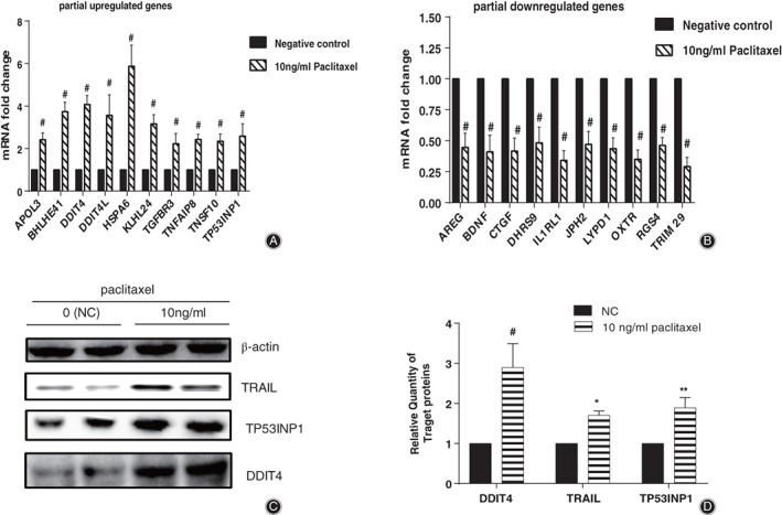
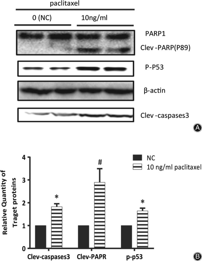
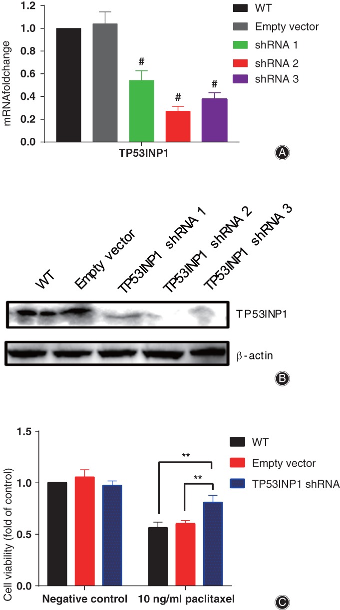
Our data revealed that paclitaxel had significant time-dependent effects on the viability and induced apoptosis of HS 737.T cells. RNA-seq and bioinformatics analysis showed that apoptosis, death receptor signaling pathway, TNF signaling pathway, and TP53 regulated transcription of cell death genes pathway were closely associated with paclitaxel in the treatment of GCTB. Western bolt results revealed that paclitaxel induced cleavage of Caspase-3 and PARP1, and increased the phosphorylation level of p53 in HS 737.T cells. RNAi results showed that the expression level of TP53INP1 was significantly decreased in HS737.T cells (the decrease was more than 70%). In addition, we found that the inhibitory ratios of paclitaxel on HS737.T cells deficient in TP53INP1 were less than in HS737.T cells with empty vector (19.88 and 40.60%, respectively). Hence, our data revealed that TP53INP1 regulated paclitaxel-driven apoptosis in HS737.T cells.
Paclitaxel can significantly repress cell proliferation and induce apoptosis of HS 737.T cells through activating Caspase-3, PARP1, p53, and TP53INP1. Paclitaxel may be an effective drug in the management of GCTB.
评估紫杉醇的抗肿瘤能力并探究其潜在分子机制。
首先,采用cck-8和凋亡检测法来确定紫杉醇处理下HS 737.T细胞的存活和凋亡效应。其次,运用RNA测序和生物信息学方法来确定差异表达基因并分析相关通路。采用定量实时聚合酶链反应来验证部分差异表达基因(DEG)的准确性。利用ClueGO对差异表达基因的功能分组GO术语进行解码和可视化,并绘制DEG蛋白-蛋白相互作用(PPI)网络。通过蛋白质免疫印迹法检测靶基因的表达、半胱天冬酶-3(Caspase-3)和聚(ADP-核糖)聚合酶1(PARP1)的裂解以及p53的磷酸化水平。最后,运用转录组学、生物信息学和RNA干扰技术来评估紫杉醇在骨巨细胞瘤(GCTB)中的抗肿瘤能力并确定其潜在机制。
我们的数据显示,紫杉醇对HS 737.T细胞的活力具有显著的时间依赖性影响并诱导其凋亡。RNA测序和生物信息学分析表明,凋亡、死亡受体信号通路、肿瘤坏死因子(TNF)信号通路以及TP53调控的细胞死亡基因转录通路与紫杉醇治疗GCTB密切相关。蛋白质免疫印迹结果显示,紫杉醇诱导HS 737.T细胞中Caspase-3和PARP1的裂解,并提高p53的磷酸化水平。RNA干扰结果表明,TP53诱导蛋白1(TP53INP1)在HS737.T细胞中的表达水平显著降低(降低超过70%)。此外,我们发现,紫杉醇对缺乏TP53INP1的HS737.T细胞的抑制率低于对空载载体HS737.T细胞的抑制率(分别为19.88%和40.60%)。因此,我们的数据表明,TP53INP1调节紫杉醇驱动的HS737.T细胞凋亡。
紫杉醇可通过激活Caspase-3、PARP1、p53和TP53INP1显著抑制HS 737.T细胞增殖并诱导其凋亡。紫杉醇可能是治疗GCTB的一种有效药物。