Jo Janggun, Tian Chao, Xu Guan, Sarazin Jeffrey, Schiopu Elena, Gandikota Girish, Wang Xueding
Department of Biomedical Engineering, University of Michigan, Ann Arbor, MI 48109, United States.
College of Engineering Science, University of Science and Technology of China, Hefei, Anhui 230026, China.
Photoacoustics. 2018 Jul 27;12:82-89. doi: 10.1016/j.pacs.2018.07.004. eCollection 2018 Dec.
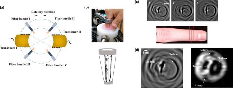
With the capability of assessing high resolution optical contrast in soft tissues, photoacoustic imaging (PAI) can offer valuable structural and functional information of human joints, and hold potential for diagnosis and treatment monitoring of inflammatory arthritis. Recent studies have demonstrated that PAI can map 2D and 3D morphology of the cartilage, synovium, vascularity, and bone tissue in human peripheral joints. Initial trials with patients affected by inflammatory arthritis have also suggested that PAI can detect the hemodynamic properties in articular tissues as well as their changes due to active inflammation. This review focuses on the recent progress in technical development of PAI for human musculoskeletal imaging and inflammation detection. PAI can provide non-invasive and non-ionizing serial measurements for monitoring of therapeutic interventions with the potential for higher sensitivity than existing imaging modalities such as ultrasound. However, further investigation is needed to validate the value of PAI in rheumatology clinical settings.
光声成像(PAI)能够评估软组织中的高分辨率光学对比度,可为人体关节提供有价值的结构和功能信息,并在炎症性关节炎的诊断和治疗监测方面具有潜力。最近的研究表明,PAI可以绘制人体外周关节中软骨、滑膜、血管和骨组织的二维和三维形态。对炎症性关节炎患者的初步试验也表明,PAI可以检测关节组织中的血液动力学特性以及由于活动性炎症引起的变化。这篇综述重点关注用于人体肌肉骨骼成像和炎症检测的PAI技术发展的最新进展。PAI可以提供非侵入性和非电离的连续测量,用于监测治疗干预措施,其灵敏度可能高于超声等现有成像方式。然而,需要进一步研究来验证PAI在风湿病临床环境中的价值。