Gonzalez-Calle Alejandra, Brant Rodrigo, Diniz Bruno, Swenson Steven, Markland Frank, Humayun Mark S, Weiland James D
Dr Allen and Charlotte Ginsburg Institute for Biomedical Therapeutics, USC Roski Eye Institute, University of Southern California, United States.
Department of Ophthalmology and Visual Sciences, Federal University of Sao Paulo, Brazil.
J Clin Exp Ophthalmol. 2018;9(5). doi: 10.4172/2155-9570.1000752. Epub 2018 Oct 8.
We propose a novel attachment method for retinal tissue that utilizes silicone modified with bioactive molecules.
This is an experimental study divided into an section performed in cadaveric pig eyes and an section performed in rabbits.
During experiments 36 cadaveric pig eyes were used. During experiments 4 rabbits were used.
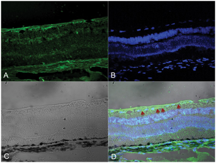
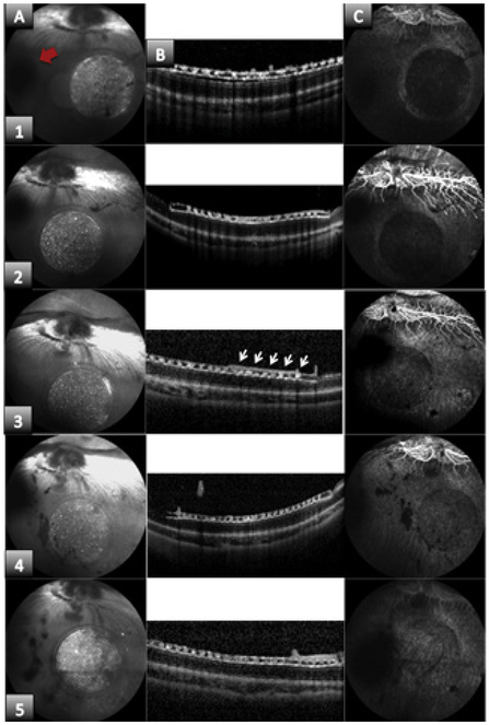
Different types of silicone went through a laser irradiation process to determine if binding sites for disintegrins could be created. Laser treated silicones that showed disintegrin binding were evaluated with testing for retina-silicone attachment. The best silicone binding was implanted into a rabbit's eye after a full vitrectomy was performed. Post-operative exams were done every two weeks to evaluate placement, attachment and sterilization method. After three months animals were euthanized and eye was enucleated for histology analysis.
Attachment strength between silicone-disintegrin-retina, and signs of endophthalmitis during studies for biocompatibility purposes.
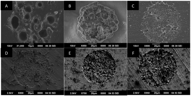
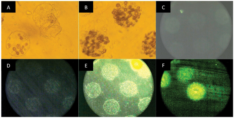
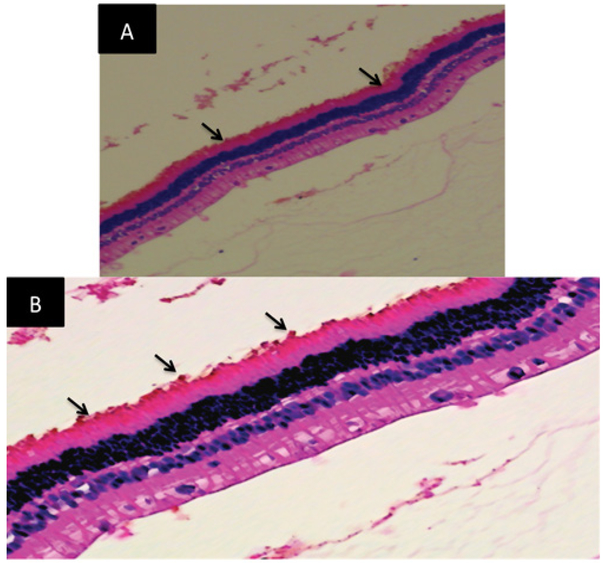
A technique to successfully lase and produce an active area on the silicone surface was described. Scanning electron microscope (SEM) images were evaluated to assess physical ablation/debris field area on the surface, definition of edges, evenness, and symmetry of the lased area allowing us to select MED 4800 silicone family for further testing. Cell culture experiments showed disintegrin binding to the silicone active area. experiments with cadaveric eyes were performed to test retina-silicone attachment. MED 4860 showed strongest attachment to the retina and it was used during experiments. A sterilization protocol was tested and proved to be reliable for bioactive materials. Disintegrin coated silicone showed attachment in 2 of 4 rabbits during the 3-month implant period. The adhesion was persistent until reversed with plasmin. All rabbits were implanted for 3 months regardless of attachment, to test the feasibility of the sterilization method. None of the rabbits developed any type of eye infection during the implant period.
We successfully lased and produced an active area on the silicone surface to allow disintegrin-silicone binding. Different silicones interact differently with the laser energy, and this is reflected in the strength of the silicone-disintegrin-retina attachment.
我们提出一种用于视网膜组织的新型附着方法,该方法利用经生物活性分子修饰的硅胶。
这是一项实验研究,分为在猪尸体眼上进行的部分和在兔子身上进行的部分。
实验过程中使用了36只猪尸体眼。实验过程中使用了4只兔子。
不同类型的硅胶经过激光照射过程,以确定是否可以产生整合素的结合位点。对显示整合素结合的经激光处理的硅胶进行视网膜 - 硅胶附着的测试评估。在进行完全玻璃体切除术后,将具有最佳硅胶结合力的材料植入兔眼。术后每两周进行一次检查,以评估植入物的位置、附着情况和灭菌方法。三个月后对动物实施安乐死并摘除眼球进行组织学分析。
硅胶 - 整合素 - 视网膜之间的附着强度,以及出于生物相容性目的在研究期间眼内炎的迹象。
描述了一种在硅胶表面成功进行激光照射并产生活性区域的技术。通过扫描电子显微镜(SEM)图像评估表面的物理消融/碎片场区域、边缘清晰度、均匀度和激光照射区域的对称性,这使我们能够选择MED 4800硅胶系列进行进一步测试。细胞培养实验表明整合素与硅胶活性区域结合。在猪尸体眼上进行实验以测试视网膜 - 硅胶附着情况。MED 4860显示出与视网膜的最强附着,并且在实验中使用。测试了一种灭菌方案,证明其对生物活性材料可靠。在3个月的植入期内,整合素包被的硅胶在4只兔子中的2只中显示出附着。这种粘附持续存在,直到用纤溶酶使其逆转。无论附着情况如何,所有兔子都植入3个月,以测试灭菌方法的可行性。在植入期内,没有兔子发生任何类型的眼部感染。
我们成功地在硅胶表面进行激光照射并产生活性区域,以实现整合素 - 硅胶结合。不同的硅胶与激光能量的相互作用不同,这反映在硅胶 - 整合素 - 视网膜附着的强度上。