Cambridge Centre for Medical Materials, Department of Materials Science and Metallurgy, University of Cambridge, Cambridge, United Kingdom.
Imaging Facility, Gurdon Institute, University of Cambridge, Cambridge, United Kingdom.
PLoS One. 2019 Jan 8;14(1):e0210390. doi: 10.1371/journal.pone.0210390. eCollection 2019.
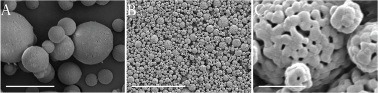
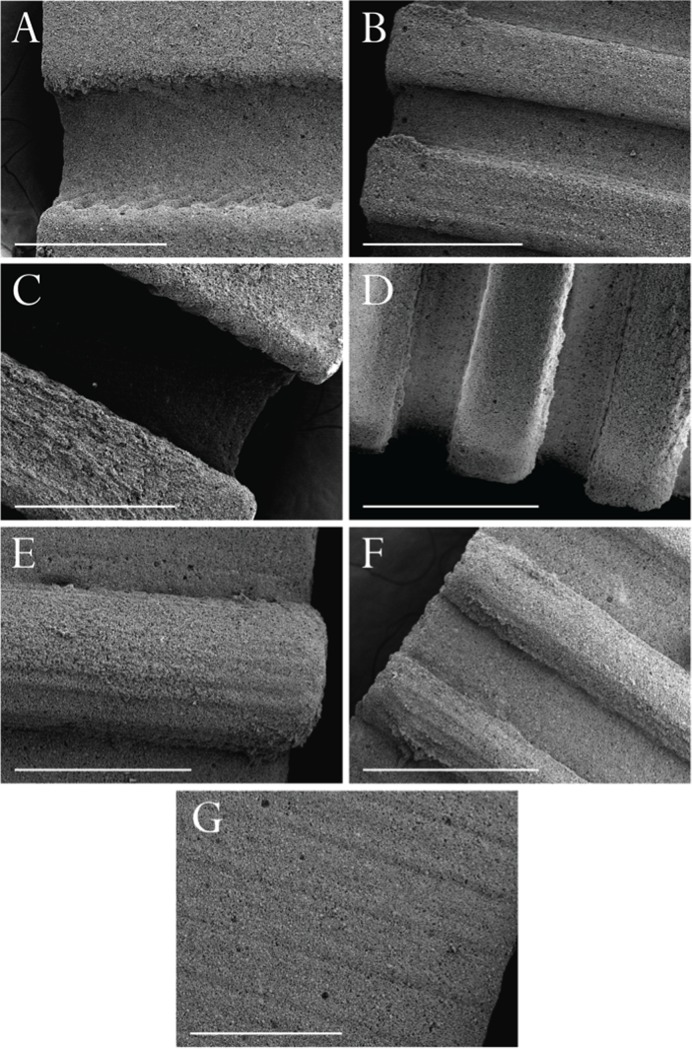
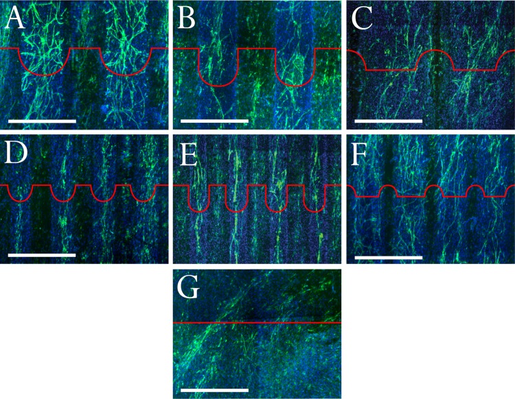
One of the greatest obstacles to clinical translation of bone tissue engineering is the inability to effectively and efficiently vascularize scaffolds. The goal of this work was to explore systematically whether architecture, at a scale of hundreds of microns, can be used to direct the growth of microcapillary-like structures into the core of scaffolds. Biphasic bioceramic patterned architectures were produced using silicone molds of 3D printed parts. Grooves and ridges were designed to have widths of 330 μm and 660 μm, with periodicities respectively of 1240 μm and 630 μm. Groove depth was varied between 150 μm and 585 μm. Co-cultures of human dermal microvascular endothelial cells (HDMECs) and human osteoblasts (hOBs) were used to grow microcapillary-like structures on substrates. Bioceramic architecture was found to significantly affect microcapillary-like structure location and orientation. Microcapillary-like structures were found to form predominantly in grooves or between convexities. For all patterned samples, the CD31 (endothelial cell marker) signal was at least 2.5 times higher along grooves versus perpendicular to grooves. In addition, the average signal was at least two times higher within grooves than outside grooves for all samples. Grooves with a width of 330 μm and a depth of 300 μm resulted in the formation of individual, highly aligned microcapillary-like structures with lengths around 5 mm. Extensive literature has focused on the role of nano- and micro-topography (on the scale below tens of microns) on cellular response. However, the idea that architecture at a scale much larger than a cell could be used to modulate angiogenesis has not been systematically investigated. This work shows the crucial influence of architecture on microcapillary-like structure self-assembly at the scale of hundreds of microns. Elucidating the precise correspondence between architecture and microcapillary-like structure organization will ultimately allow the engineering of microvasculature by tuning local scaffold design to achieve desirable microvessel properties.
组织工程学中骨组织临床转化的最大障碍之一是无法有效地使支架血管化。本工作的目的是系统地探索在数百微米的尺度上,支架的结构能否用于引导类毛细血管样结构向支架内部生长。使用 3D 打印零件的硅模具制作双相生物陶瓷图案化结构。设计了宽度分别为 330μm 和 660μm 的沟槽和脊,其周期分别为 1240μm 和 630μm。沟槽深度在 150μm 和 585μm 之间变化。将人真皮微血管内皮细胞(HDMEC)和人成骨细胞(hOB)共培养物用于在基底上生长类毛细血管样结构。发现生物陶瓷结构显著影响类毛细血管样结构的位置和取向。发现类毛细血管样结构主要在沟槽或凸部之间形成。对于所有图案化样品,与垂直于沟槽相比,沟槽处的 CD31(内皮细胞标志物)信号至少高 2.5 倍。此外,对于所有样品,沟槽内的平均信号至少是沟槽外的两倍。宽度为 330μm 且深度为 300μm 的沟槽导致形成具有 5mm 左右长度的单个、高度对齐的类毛细血管样结构。大量文献集中研究了纳米和微观形貌(在数十微米以下的尺度上)对细胞反应的作用。然而,用于调节血管生成的结构比细胞大得多的尺度的想法尚未被系统地研究。这项工作表明了结构对数百微米尺度类毛细血管样结构自组装的关键影响。阐明结构与类毛细血管样结构组织之间的确切对应关系将最终允许通过调整局部支架设计来实现理想的微血管特性来工程化微血管。