Department of Biomaterials, Max Planck Institute of Colloids and Interfaces, Potsdam, Germany.
PLoS One. 2012;7(5):e36336. doi: 10.1371/journal.pone.0036336. Epub 2012 May 11.
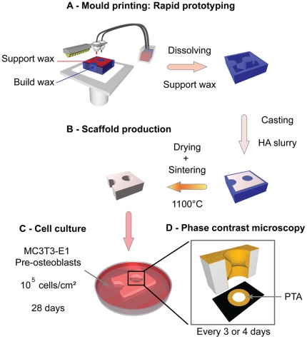
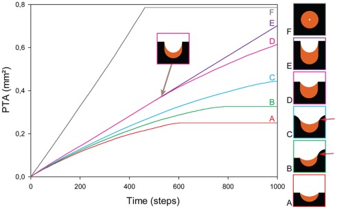
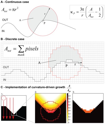
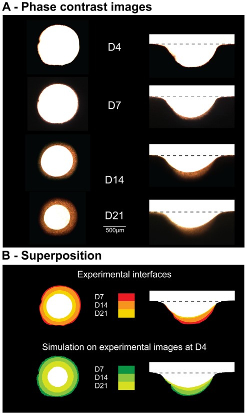
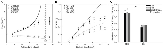
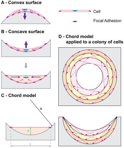
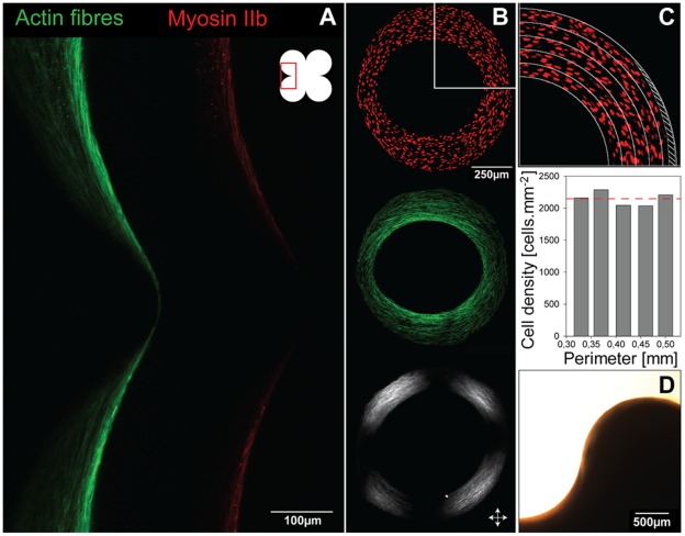
This study investigated how substrate geometry influences in-vitro tissue formation at length scales much larger than a single cell. Two-millimetre thick hydroxyapatite plates containing circular pores and semi-circular channels of 0.5 mm radius, mimicking osteons and hemi-osteons respectively, were incubated with MC3T3-E1 cells for 4 weeks. The amount and shape of the tissue formed in the pores, as measured using phase contrast microscopy, depended on the substrate geometry. It was further demonstrated, using a simple geometric model, that the observed curvature-controlled growth can be derived from the assembly of tensile elements on a curved substrate. These tensile elements are cells anchored on distant points of the curved surface, thus creating an actin "chord" by generating tension between the adhesion sites. Such a chord model was used to link the shape of the substrate to cell organisation and tissue patterning. In a pore with a circular cross-section, tissue growth increases the average curvature of the surface, whereas a semi-circular channel tends to be flattened out. Thereby, a single mechanism could describe new tissue growth in both cortical and trabecular bone after resorption due to remodelling. These similarities between in-vitro and in-vivo patterns suggest geometry as an important signal for bone remodelling.
本研究探讨了在远大于单个细胞的长度尺度上,基底几何形状如何影响体外组织的形成。将含有 0.5 毫米半径的圆形孔和半圆形通道的 2 毫米厚的羟基磷灰石板分别模拟成骨单位和半骨单位,与 MC3T3-E1 细胞共培养 4 周。通过相差显微镜测量孔内形成的组织的数量和形状,发现其取决于基底的几何形状。进一步通过一个简单的几何模型证明,观察到的曲率控制生长可以从在弯曲基底上组装拉伸元件中得出。这些拉伸元件是在弯曲表面的远处固定的细胞,通过在附着点之间产生张力,从而形成一个肌动蛋白“弦”。这样的弦模型被用来将基底的形状与细胞组织和组织模式联系起来。在具有圆形横截面的孔中,组织生长会增加表面的平均曲率,而半圆形通道则趋于变平。因此,一种单一的机制可以描述由于重塑而在皮质骨和小梁骨中吸收后新组织的生长。这种体外和体内模式之间的相似性表明,几何形状是骨骼重塑的一个重要信号。