Department of Medical Imaging and Intervention, Imaging Core Laboratory, Institute for Radiological Research, Chang Gung Memorial Hospital at Linkou, Taoyuan, Taiwan.
Department of Diagnostic Radiology, Chang Gung Memorial Hospital at Keelung, Keelung, Taiwan.
Korean J Radiol. 2019 Jan;20(1):18-33. doi: 10.3348/kjr.2018.0090. Epub 2018 Dec 27.
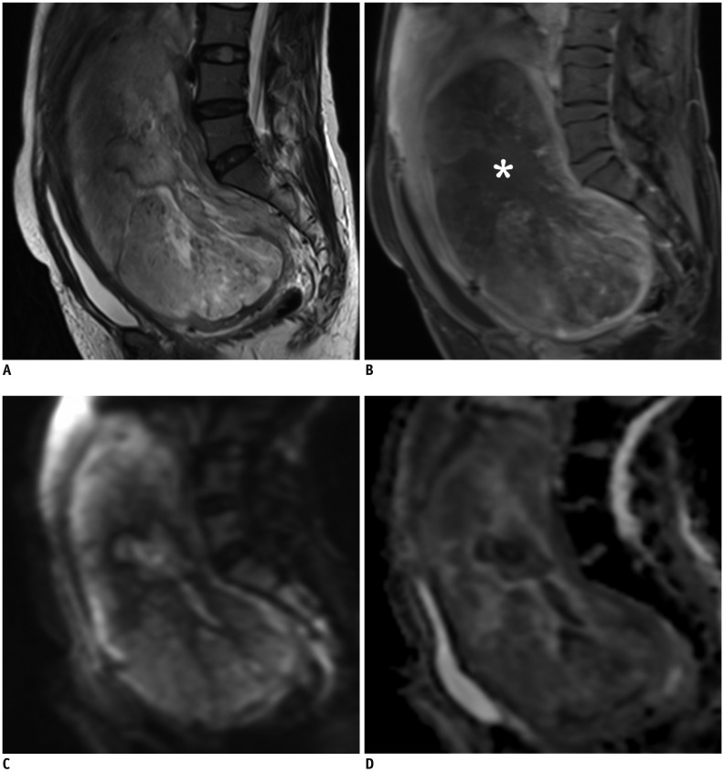
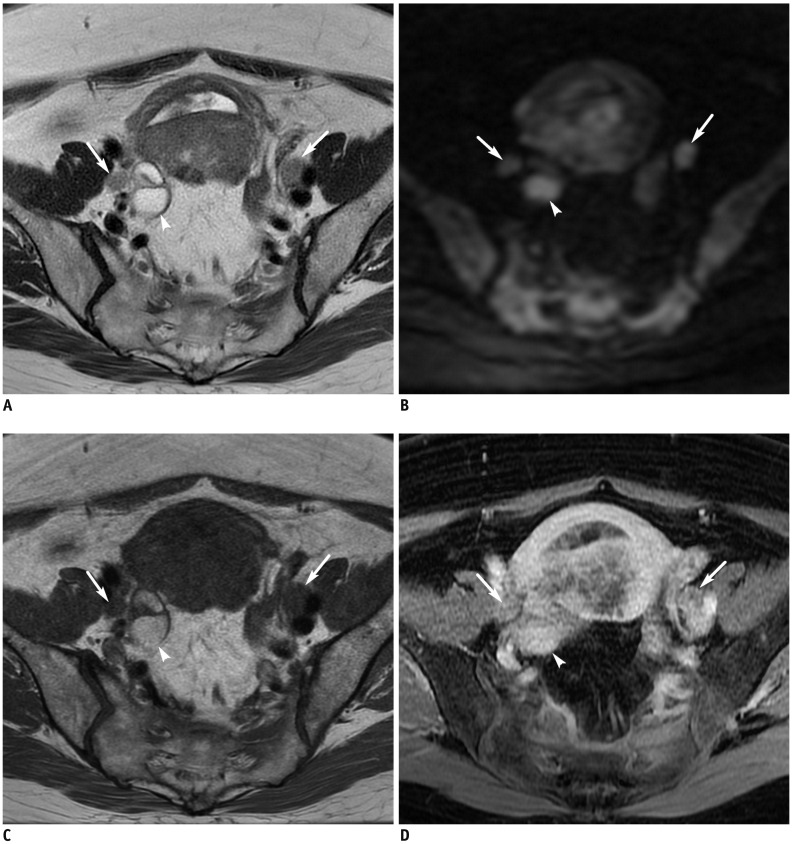
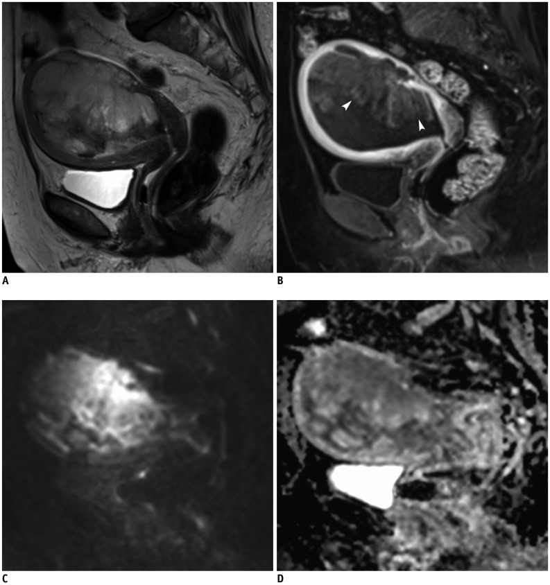
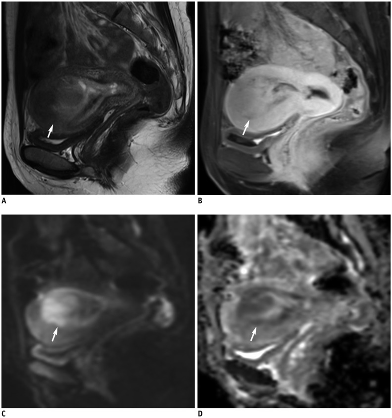
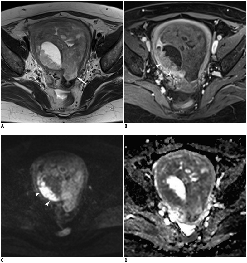
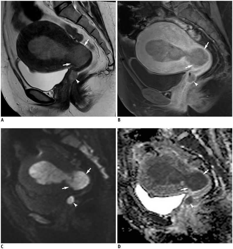
In this study, we summarize the clinical role of magnetic resonance imaging (MRI) in the diagnosis of patients with malignant uterine neoplasms, including leiomyosarcoma, endometrial stromal sarcoma, adenosarcoma, uterine carcinosarcoma, and endometrial cancer, with emphasis on the challenges and disadvantages. MRI plays an essential role in patients with uterine malignancy, for the purpose of tumor detection, primary staging, and treatment planning. MRI has advanced in scope beyond the visualization of the many aspects of anatomical structures, including diffusion-weighted imaging, dynamic contrast enhancement-MRI, and magnetic resonance spectroscopy. Emerging technologies coupled with the use of artificial intelligence in MRI are expected to lead to progressive improvement in case management of malignant uterine neoplasms.
在这项研究中,我们总结了磁共振成像(MRI)在诊断恶性子宫肿瘤患者中的临床作用,包括平滑肌肉瘤、子宫内膜间质肉瘤、腺肉瘤、子宫癌肉瘤和子宫内膜癌,重点介绍了其挑战和缺点。MRI 在子宫恶性肿瘤患者中具有重要作用,可用于肿瘤检测、原发分期和治疗计划。MRI 的应用范围已经超越了对解剖结构多个方面的可视化,包括扩散加权成像、动态对比增强 MRI 和磁共振波谱。新兴技术与 MRI 中人工智能的使用相结合,有望逐步改善恶性子宫肿瘤的病例管理。