Department of Clinical Medicine, Laboratory of Experimental Therapeutics, School of Medicine, University of Sao Paulo, Sao Paulo, Brazil.
Department of Pathology, Laboratory of Molecular Pathology, School of Medicine, University of Sao Paulo, Sao Paulo, Brazil.
PLoS One. 2019 Jan 10;14(1):e0209351. doi: 10.1371/journal.pone.0209351. eCollection 2019.
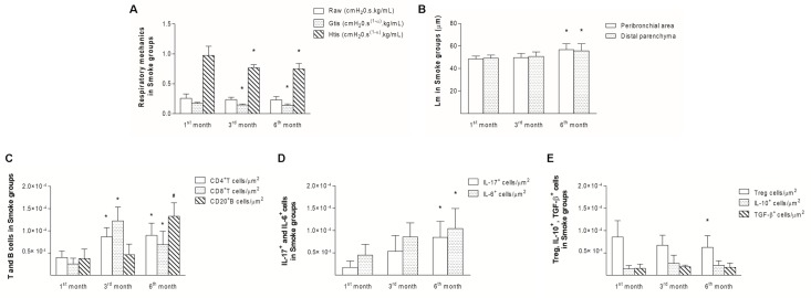
The imbalance between pro- and anti-inflammatory immune responses plays a pivotal role in chronic obstructive pulmonary disease (COPD) development and progression. To clarify the pathophysiological mechanisms of this disease, we performed a temporal analysis of immune response-mediated inflammatory progression in a cigarette smoke (CS)-induced mouse model with a focus on the balance between Th17 and Treg responses.
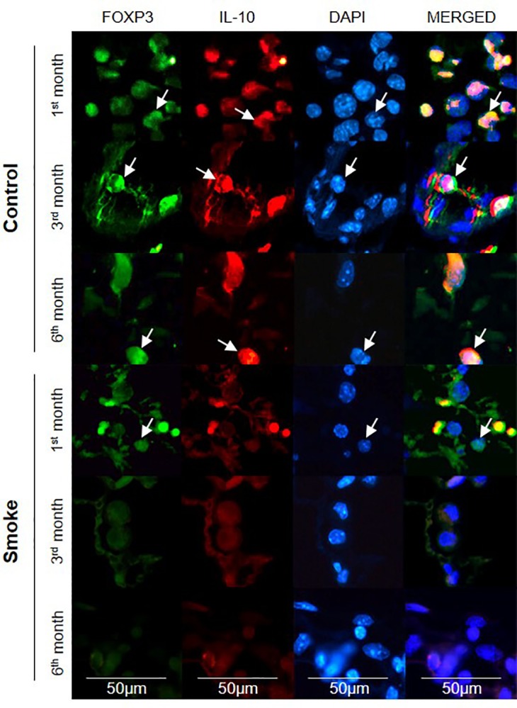
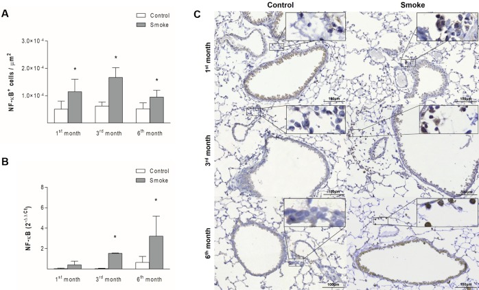
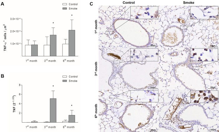
C57BL/6 mice were exposed to CS for 1, 3 or 6 months to induce COPD, and the control groups were maintained under filtered air conditions for the same time intervals. We then performed functional (respiratory mechanics) and structural (alveolar enlargement) analyses. We also quantified the NF-κB, TNF-α, CD4, CD8, CD20, IL-17, IL-6, FOXP3, IL-10, or TGF-β positive cells in peribronchovascular areas and assessed FOXP3 and IL-10 expression through double-label immunofluorescence. Additionally, we evaluated the gene expression of NF-κB and TNF in bronchiolar epithelial cells.
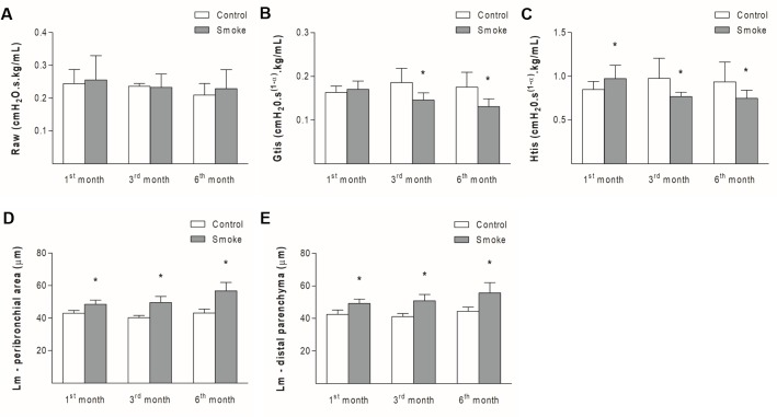
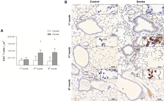
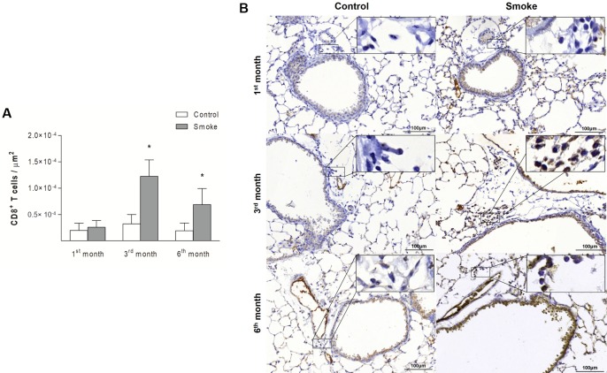
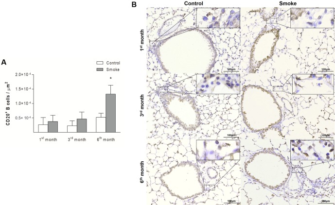
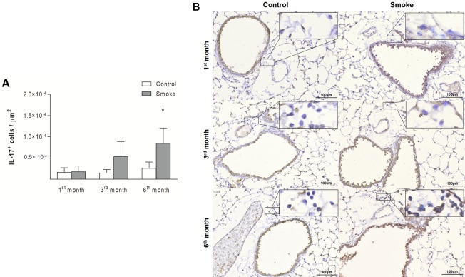
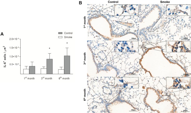
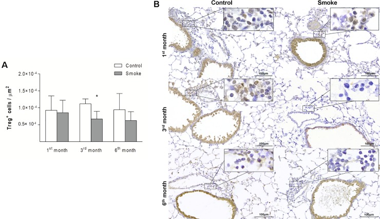
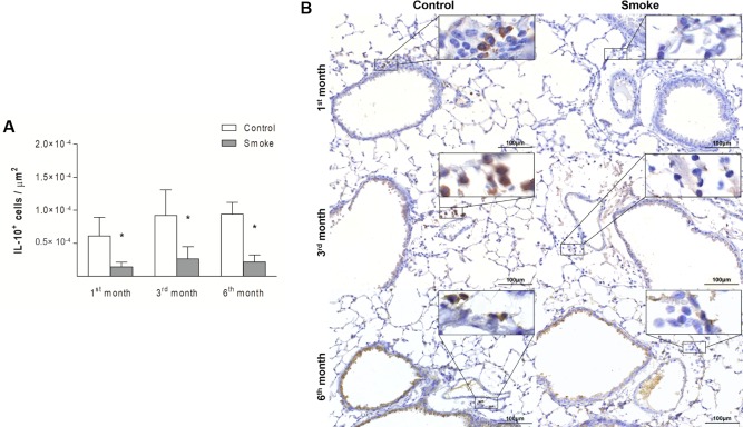
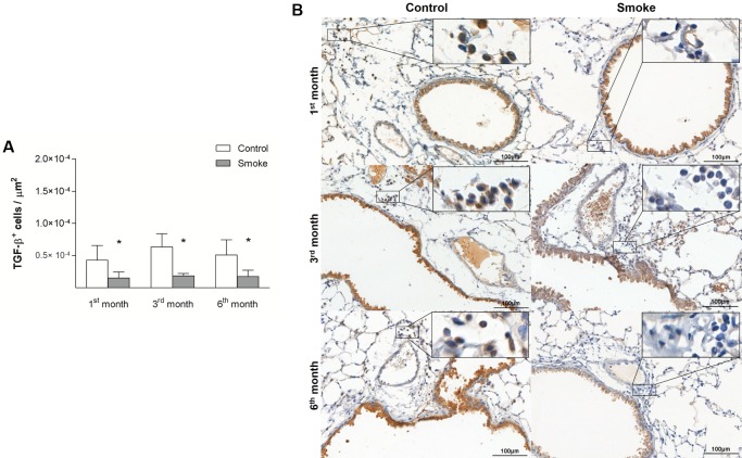
Our CS-induced COPD model exhibited an increased proinflammatory immune response (increased expression of the NF-κB, TNF-α, CD4, CD8, CD20, IL-17, and IL-6 markers) with a concomitantly decreased anti-inflammatory immune response (FOXP3, IL-10, and TGF-β markers) compared with the control mice. These changes in the immune responses were associated with increased alveolar enlargement and impaired lung function starting on the first month and third month of CS exposure, respectively, compared with the control mice.
Our results showed that the microenvironmental stimuli produced by the release of cytokines during COPD progression lead to a Th17/Treg imbalance.
促炎和抗炎免疫反应失衡在慢性阻塞性肺疾病(COPD)的发展和进展中起着关键作用。为了阐明这种疾病的病理生理机制,我们在香烟烟雾(CS)诱导的小鼠模型中进行了免疫反应介导的炎症进展的时间分析,重点关注 Th17 和 Treg 反应之间的平衡。
C57BL/6 小鼠暴露于 CS 中 1、3 或 6 个月以诱导 COPD,对照组在相同的时间间隔内保持在过滤空气中。然后,我们进行了功能(呼吸力学)和结构(肺泡扩大)分析。我们还定量了 NF-κB、TNF-α、CD4、CD8、CD20、IL-17、IL-6、FOXP3、IL-10 或 TGF-β在支气管血管周围区域的阳性细胞,并通过双标免疫荧光评估 FOXP3 和 IL-10 的表达。此外,我们评估了 NF-κB 和 TNF 在细支气管上皮细胞中的基因表达。
与对照组相比,我们的 CS 诱导的 COPD 模型表现出促炎免疫反应增加(NF-κB、TNF-α、CD4、CD8、CD20、IL-17 和 IL-6 标志物表达增加),同时抗炎免疫反应减少(FOXP3、IL-10 和 TGF-β 标志物)。与对照组相比,这些免疫反应的变化与暴露于 CS 后的第一个月和第三个月分别开始的肺泡扩大和肺功能受损有关。
我们的结果表明,在 COPD 进展过程中细胞因子释放产生的微环境刺激导致 Th17/Treg 失衡。