• 文献检索
  • 文档翻译
  • 深度研究
  • 学术资讯
  • Suppr Zotero 插件Zotero 插件
  • 邀请有礼
  • 套餐&价格
  • 历史记录
应用&插件
Suppr Zotero 插件Zotero 插件浏览器插件Mac 客户端Windows 客户端微信小程序
定价
高级版会员购买积分包购买API积分包
服务
文献检索文档翻译深度研究API 文档MCP 服务
关于我们
关于 Suppr公司介绍联系我们用户协议隐私条款
关注我们

Suppr 超能文献

核心技术专利:CN118964589B侵权必究
粤ICP备2023148730 号-1Suppr @ 2026

文献检索

告别复杂PubMed语法,用中文像聊天一样搜索,搜遍4000万医学文献。AI智能推荐,让科研检索更轻松。

立即免费搜索

文件翻译

保留排版,准确专业,支持PDF/Word/PPT等文件格式,支持 12+语言互译。

免费翻译文档

深度研究

AI帮你快速写综述,25分钟生成高质量综述,智能提取关键信息,辅助科研写作。

立即免费体验

哪种中央型肿瘤的定义对于影像学 N0 期非小细胞肺癌患者隐匿性纵隔转移的预测更有价值?

Which definition of a central tumour is more predictive of occult mediastinal metastasis in nonsmall cell lung cancer patients with radiological N0 disease?

机构信息

Division of Pulmonary and Critical Care Medicine, Dept of Medicine, Samsung Medical Center, Sungkyunkwan University School of Medicine, Seoul, Republic of Korea.

These two authors contributed equally to this work.

出版信息

Eur Respir J. 2019 Mar 18;53(3). doi: 10.1183/13993003.01508-2018. Print 2019 Mar.

DOI:10.1183/13993003.01508-2018
PMID:30635291
原文链接:https://pmc.ncbi.nlm.nih.gov/articles/PMC6422838/
Abstract

BACKGROUND

Guidelines recommend invasive mediastinal staging for centrally located tumours, even in radiological N0 nonsmall cell lung cancer (NSCLC). However, there is no uniform definition of a central tumour that is more predictive of occult mediastinal metastasis.

METHODS

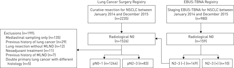
A total of 1337 consecutive patients with radiological N0 disease underwent invasive mediastinal staging. Tumours were categorised into central and peripheral by seven different definitions.

RESULTS

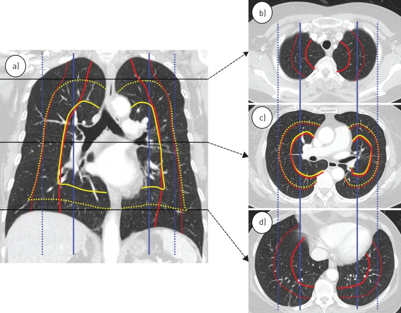
About 7% (93 out of 1337) of patients had occult N2 disease, and they had significantly larger tumour size and more solid tumours on computed tomography. After adjustment for patient- and tumour-related characteristics, only the central tumour definition of the inner one-third of the hemithorax adopted by drawing concentric lines arising from the midline significantly predicted occult N2 disease (adjusted OR 2.13, 95% CI 1.17-3.87; p=0.013). This association was maintained after excluding patients with pure ground-glass nodules (adjusted OR 2.54, 95% CI 1.37-4.71; p=0.003) or only including those with solid tumours (adjusted OR 2.30, 95% CI 1.08-4.88; p=0.030).

CONCLUSIONS

We suggest that a central tumour should be defined using the inner one-third of the hemithorax adopted by drawing concentric lines from the midline. This is particularly useful for predicting occult N2 disease in patients with NSCLC.

摘要

背景

指南建议对中央型肿瘤进行有创性纵隔分期,即使在影像学 N0 非小细胞肺癌(NSCLC)中也是如此。然而,对于更能预测隐匿性纵隔转移的中央型肿瘤,目前尚无统一的定义。

方法

共对 1337 例影像学 N0 疾病的连续患者进行了有创性纵隔分期。肿瘤通过七种不同的定义分为中央型和周围型。

结果

约 7%(93/1337)的患者存在隐匿性 N2 疾病,且其肿瘤体积更大,CT 上实性肿瘤更多。在调整了患者和肿瘤相关特征后,仅从中线引出的同心线将胸腔的内三分之一作为中央型肿瘤定义,能显著预测隐匿性 N2 疾病(调整后的 OR 2.13,95%CI 1.17-3.87;p=0.013)。在排除了纯磨玻璃结节(调整后的 OR 2.54,95%CI 1.37-4.71;p=0.003)或仅包括实性肿瘤的患者(调整后的 OR 2.30,95%CI 1.08-4.88;p=0.030)后,这种相关性仍然存在。

结论

我们建议采用从中线引出的同心线将胸腔的内三分之一作为中央型肿瘤的定义。这对于预测 NSCLC 患者隐匿性 N2 疾病特别有用。

https://cdn.ncbi.nlm.nih.gov/pmc/blobs/ee6a/6422838/71355f205c67/ERJ-01508-2018.03.jpg
https://cdn.ncbi.nlm.nih.gov/pmc/blobs/ee6a/6422838/99e303b2de6f/ERJ-01508-2018.01.jpg
https://cdn.ncbi.nlm.nih.gov/pmc/blobs/ee6a/6422838/f113f720f5d7/ERJ-01508-2018.02.jpg
https://cdn.ncbi.nlm.nih.gov/pmc/blobs/ee6a/6422838/71355f205c67/ERJ-01508-2018.03.jpg
https://cdn.ncbi.nlm.nih.gov/pmc/blobs/ee6a/6422838/99e303b2de6f/ERJ-01508-2018.01.jpg
https://cdn.ncbi.nlm.nih.gov/pmc/blobs/ee6a/6422838/f113f720f5d7/ERJ-01508-2018.02.jpg
https://cdn.ncbi.nlm.nih.gov/pmc/blobs/ee6a/6422838/71355f205c67/ERJ-01508-2018.03.jpg

相似文献

1
Which definition of a central tumour is more predictive of occult mediastinal metastasis in nonsmall cell lung cancer patients with radiological N0 disease?哪种中央型肿瘤的定义对于影像学 N0 期非小细胞肺癌患者隐匿性纵隔转移的预测更有价值?
Eur Respir J. 2019 Mar 18;53(3). doi: 10.1183/13993003.01508-2018. Print 2019 Mar.
2
Centrally located lung cancer and risk of occult nodal disease: an objective evaluation of multiple definitions of tumour centrality with dedicated imaging software.中央型肺癌与隐匿性淋巴结疾病风险:利用专用成像软件对肿瘤中央性的多种定义进行客观评估。
Eur Respir J. 2019 May 9;53(5). doi: 10.1183/13993003.02220-2018. Print 2019 May.
3
Is central lung tumour location really predictive for occult mediastinal nodal disease in (suspected) non-small-cell lung cancer staged cN0 on 18F-fluorodeoxyglucose positron emission tomography-computed tomography?中央肺部肿瘤位置对于 18F-氟脱氧葡萄糖正电子发射断层扫描-计算机断层扫描分期为 cN0(疑似)非小细胞肺癌的隐匿性纵隔淋巴结疾病是否具有真正的预测价值?
Eur J Cardiothorac Surg. 2018 Jul 1;54(1):134-140. doi: 10.1093/ejcts/ezy018.
4
Pattern and predictors of occult mediastinal lymph node involvement in non-small cell lung cancer patients with negative mediastinal uptake on positron emission tomography.正电子发射断层扫描显示纵隔摄取阴性的非小细胞肺癌患者隐匿性纵隔淋巴结受累的模式及预测因素
Eur J Cardiothorac Surg. 2008 Jan;33(1):104-9. doi: 10.1016/j.ejcts.2007.09.026. Epub 2007 Oct 30.
5
Association of maximum standardized uptake value with occult mediastinal lymph node metastases in cN0 non-small cell lung cancer.cN0 期非小细胞肺癌中最大标准化摄取值与隐匿性纵隔淋巴结转移的相关性
Eur J Cardiothorac Surg. 2016 Nov;50(5):914-919. doi: 10.1093/ejcts/ezw109. Epub 2016 Apr 24.
6
Indications for invasive mediastinal staging in patients with early non-small cell lung cancer staged with PET-CT.经PET-CT分期的早期非小细胞肺癌患者进行有创纵隔分期的指征。
Lung Cancer. 2017 Jul;109:36-41. doi: 10.1016/j.lungcan.2017.04.018. Epub 2017 Apr 25.
7
Combined Endosonographic Mediastinal Lymph Node Staging in Positron Emission Tomography and Computed Tomography Node-Negative Non-Small-Cell Lung Cancer in High-Risk Patients.在高危患者中,正电子发射断层扫描和计算机断层扫描淋巴结阴性非小细胞肺癌的联合超声内镜纵隔淋巴结分期。
Semin Thorac Cardiovasc Surg. 2020 Spring;32(1):162-168. doi: 10.1053/j.semtcvs.2019.07.007. Epub 2019 Jul 17.
8
Prognostic value of primary tumor FDG uptake for occult mediastinal lymph node involvement in clinically N2/N3 node-negative non-small cell lung cancer.原发性肿瘤FDG摄取对临床N2/N3淋巴结阴性的非小细胞肺癌隐匿性纵隔淋巴结受累的预后价值
Am J Clin Oncol. 2014 Apr;37(2):135-9. doi: 10.1097/COC.0b013e31826b9cd3.
9
Definitions of Central Tumors in Radiologically Node-Negative, Early-Stage Lung Cancer for Preoperative Mediastinal Lymph Node Staging: A Dual-Institution, Multireader Study.影像学检查淋巴结阴性的早期肺癌术前纵隔淋巴结分期的中央型肿瘤定义:一项双机构、多读者研究。
Chest. 2022 May;161(5):1393-1406. doi: 10.1016/j.chest.2021.11.005. Epub 2021 Nov 14.
10
False-negative rate after positron emission tomography/computer tomography scan for mediastinal staging in cI stage non-small-cell lung cancer.cI 期非小细胞肺癌纵隔分期正电子发射断层扫描/计算机断层扫描的假阴性率。
Eur J Cardiothorac Surg. 2012 Jul;42(1):93-100; discussion 100. doi: 10.1093/ejcts/ezr272. Epub 2012 Jan 20.

引用本文的文献

1
Clinical and Bronchoscopy Assessment in Diagnosing the Histopathology Type of Primary Central Lung Tumors.临床及支气管镜检查评估对原发性中央型肺肿瘤组织病理学类型的诊断价值
Open Respir Med J. 2024 Jun 6;18:e18743064318977. doi: 10.2174/0118743064318977240531100045. eCollection 2024.
2
[Impact of the Size and Depth of Pulmonary Nodules on the Surgical Approach 
for Lung Resection in the Treatment of Early-stage Lung Cancer ≤2 cm].[肺结节大小和深度对≤2cm早期肺癌肺切除手术方式的影响]
Zhongguo Fei Ai Za Zhi. 2024 Mar 20;27(3):170-178. doi: 10.3779/j.issn.1009-3419.2024.101.08.
3
Machine learning model for circulating tumor DNA detection in chronic obstructive pulmonary disease patients with lung cancer.

本文引用的文献

1
Indications for invasive mediastinal staging in patients with early non-small cell lung cancer staged with PET-CT.经PET-CT分期的早期非小细胞肺癌患者进行有创纵隔分期的指征。
Lung Cancer. 2017 Jul;109:36-41. doi: 10.1016/j.lungcan.2017.04.018. Epub 2017 Apr 25.
2
Cost-effectiveness of mediastinal lymph node staging in non-small cell lung cancer.非小细胞肺癌纵隔淋巴结分期的成本效益。
J Thorac Cardiovasc Surg. 2017 Jun;153(6):1567-1578. doi: 10.1016/j.jtcvs.2016.12.048. Epub 2017 Feb 9.
3
A Prediction Model to Help with the Assessment of Adenopathy in Lung Cancer: HAL.
用于检测慢性阻塞性肺疾病合并肺癌患者循环肿瘤DNA的机器学习模型
Transl Lung Cancer Res. 2024 Jan 31;13(1):112-125. doi: 10.21037/tlcr-23-633. Epub 2024 Jan 29.
4
Role of invasive mediastinal nodal staging in survival outcomes of patients with non-small cell lung cancer and without radiologic lymph node metastasis: a retrospective cohort study.侵袭性纵隔淋巴结分期在无放射学淋巴结转移的非小细胞肺癌患者生存结局中的作用:一项回顾性队列研究
EClinicalMedicine. 2024 Feb 10;69:102478. doi: 10.1016/j.eclinm.2024.102478. eCollection 2024 Mar.
5
Clinical Effect of Endosonography on Overall Survival in Patients with Radiological N1 Non-Small Cell Lung Cancer.超声内镜检查对影像学 N1 期非小细胞肺癌患者总生存率的临床影响。
Cancer Res Treat. 2024 Apr;56(2):502-512. doi: 10.4143/crt.2023.840. Epub 2023 Dec 4.
6
PET/CT based cross-modal deep learning signature to predict occult nodal metastasis in lung cancer.基于 PET/CT 的跨模态深度学习特征预测肺癌隐匿性淋巴结转移。
Nat Commun. 2023 Nov 18;14(1):7513. doi: 10.1038/s41467-023-42811-4.
7
Diagnostic Performance of Endosonography to Detect Mediastinal Lymph Node Metastasis in Patients with Radiological N1 Non-Small Cell Lung Cancer.超声内镜诊断影像学 N1 期非小细胞肺癌纵隔淋巴结转移的效能。
Cancer Res Treat. 2023 Jul;55(3):832-840. doi: 10.4143/crt.2022.1428. Epub 2023 Mar 2.
8
Endoscopic Technologies for Peripheral Pulmonary Lesions: From Diagnosis to Therapy.用于周围型肺病变的内镜技术:从诊断到治疗
Life (Basel). 2023 Jan 17;13(2):254. doi: 10.3390/life13020254.
9
Solitary Pulmonary Capillary Hemangioma: CT and PET-CT Features with Clinicopathologic Correlation.孤立性肺毛细血管血管瘤:CT及PET-CT特征与临床病理对照
Diagnostics (Basel). 2022 Oct 28;12(11):2618. doi: 10.3390/diagnostics12112618.
10
LobE-Specific lymph node diSsectiON for clinical early-stage non-small cell lung cancer: protocol for a randomised controlled trial (the LESSON trial).肺叶特异性淋巴结清扫术治疗临床早期非小细胞肺癌:一项随机对照试验方案(LESSON 试验)。
BMJ Open. 2022 Aug 29;12(8):e056043. doi: 10.1136/bmjopen-2021-056043.
一种用于辅助评估肺癌淋巴结病的预测模型:HAL。
Am J Respir Crit Care Med. 2017 Jun 15;195(12):1651-1660. doi: 10.1164/rccm.201607-1397OC.
4
What Exactly Is a Centrally Located Lung Tumor? Results of an Online Survey.中央型肺部肿瘤究竟是什么?一项在线调查的结果。
Ann Am Thorac Soc. 2017 Jan;14(1):118-123. doi: 10.1513/AnnalsATS.201607-568BC.
5
The International Association for the Study of Lung Cancer Lung Cancer Staging Project: Proposals for the Revision of the N Descriptors in the Forthcoming 8th Edition of the TNM Classification for Lung Cancer.国际肺癌研究协会肺癌分期项目:对即将发布的第 8 版肺癌 TNM 分类中 N 描述符修订的建议。
J Thorac Oncol. 2015 Dec;10(12):1675-84. doi: 10.1097/JTO.0000000000000678.
6
Lung cancer diagnosis and staging with endobronchial ultrasound-guided transbronchial needle aspiration compared with conventional approaches: an open-label, pragmatic, randomised controlled trial.经支气管超声引导针吸活检术与常规方法诊断和分期肺癌的比较:一项开放标签、实用、随机对照试验。
Lancet Respir Med. 2015 Apr;3(4):282-9. doi: 10.1016/S2213-2600(15)00029-6. Epub 2015 Feb 4.
7
Endobronchial ultrasound versus mediastinoscopy for mediastinal nodal staging of non-small-cell lung cancer.经支气管超声内镜与纵隔镜检查用于非小细胞肺癌纵隔淋巴结分期。
J Thorac Oncol. 2015 Feb;10(2):331-7. doi: 10.1097/JTO.0000000000000388.
8
Endobronchial ultrasound-guided transbronchial needle aspiration for systematic nodal staging of lung cancer in patients with N0 disease by computed tomography and integrated positron emission tomography-computed tomography.对于经计算机断层扫描和正电子发射断层扫描-计算机断层扫描显示为N0期疾病的肺癌患者,采用支气管内超声引导下经支气管针吸活检进行系统性淋巴结分期。
Ann Am Thorac Soc. 2015 Mar;12(3):415-9. doi: 10.1513/AnnalsATS.201409-429OC.
9
Combined endobronchial and transesophageal approach of an ultrasound bronchoscope for mediastinal staging of lung cancer.超声支气管镜联合经食管途径用于肺癌纵隔分期
PLoS One. 2014 Mar 14;9(3):e91893. doi: 10.1371/journal.pone.0091893. eCollection 2014.
10
Revised ESTS guidelines for preoperative mediastinal lymph node staging for non-small-cell lung cancer.非小细胞肺癌术前纵隔淋巴结分期的修订版欧洲胸外科医师协会指南。
Eur J Cardiothorac Surg. 2014 May;45(5):787-98. doi: 10.1093/ejcts/ezu028. Epub 2014 Feb 26.