Picower Institute for Learning and Memory, Massachusetts Institute of Technology, Cambridge, MA, 02139, USA.
Department of Biological Engineering, Massachusetts Institute of Technology, Cambridge, MA, 02139, USA.
Nat Commun. 2019 Jan 11;10(1):177. doi: 10.1038/s41467-018-08179-6.
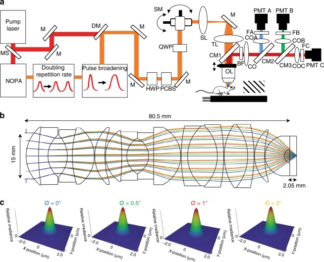
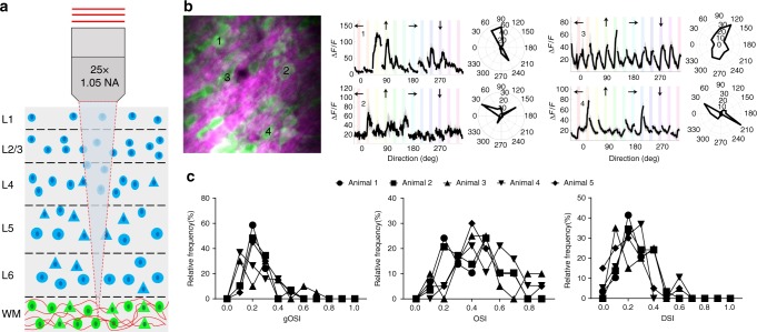
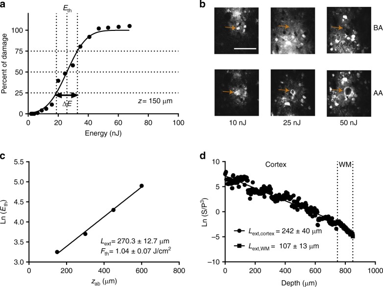
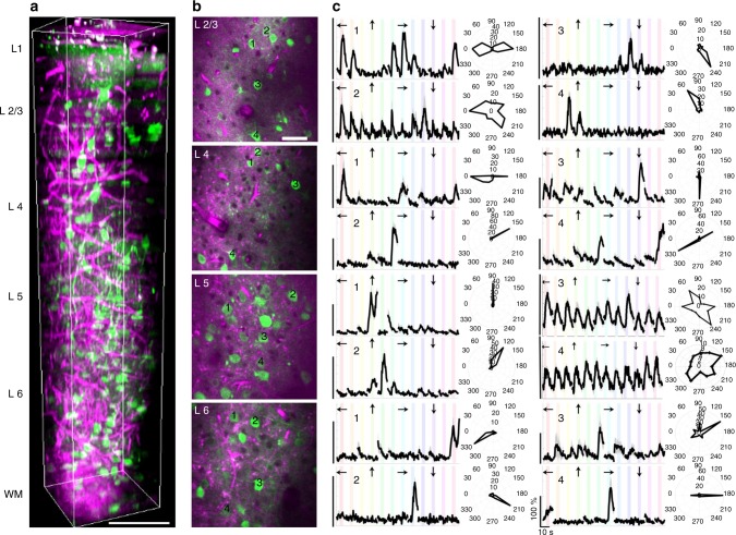
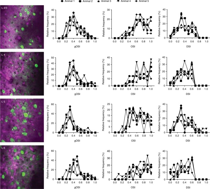
Two-photon microscopy is used to image neuronal activity, but has severe limitations for studying deeper cortical layers. Here, we developed a custom three-photon microscope optimized to image a vertical column of the cerebral cortex > 1 mm in depth in awake mice with low (<20 mW) average laser power. Our measurements of physiological responses and tissue-damage thresholds define pulse parameters and safety limits for damage-free three-photon imaging. We image functional visual responses of neurons expressing GCaMP6s across all layers of the primary visual cortex (V1) and in the subplate. These recordings reveal diverse visual selectivity in deep layers: layer 5 neurons are more broadly tuned to visual stimuli, whereas mean orientation selectivity of layer 6 neurons is slightly sharper, compared to neurons in other layers. Subplate neurons, located in the white matter below cortical layer 6 and characterized here for the first time, show low visual responsivity and broad orientation selectivity.
双光子显微镜被用于对神经元活动进行成像,但在研究较深层的皮质层时有严重的局限性。在这里,我们开发了一种定制的三光子显微镜,该显微镜经过优化,可在低(<20mW)平均激光功率下对清醒小鼠的大脑皮层中深度超过 1mm 的垂直柱进行成像。我们对生理反应和组织损伤阈值的测量定义了无损伤三光子成像的脉冲参数和安全限制。我们对表达 GCaMP6s 的神经元在初级视觉皮层(V1)的所有层以及在基板中的功能视觉反应进行成像。这些记录显示了深层层中的多种视觉选择性:与其他层中的神经元相比,第 5 层神经元对视觉刺激的调谐更广泛,而第 6 层神经元的平均方向选择性稍微更尖锐。基板神经元位于皮质第 6 层下方的白质中,这里是首次对其进行描述,它们表现出较低的视觉反应性和广泛的方向选择性。