QIMP Team, Center for Medical Physics and Biomedical Engineering, Medical University of Vienna, Vienna, Austria.
Division of Nuclear Medicine, Department of Biomedical Imaging and Image-guided Therapy, Medical University of Vienna, Waehringer Guertel 18-20, 1090, Vienna, Austria.
Eur Radiol. 2019 Aug;29(8):4276-4285. doi: 10.1007/s00330-018-5942-9. Epub 2019 Jan 11.
To assess if tumour grading based on dynamic [18F]FET positron emission tomography/magnetic resonance imaging (PET/MRI) studies is affected by different MRI-based attenuation correction (AC) methods.
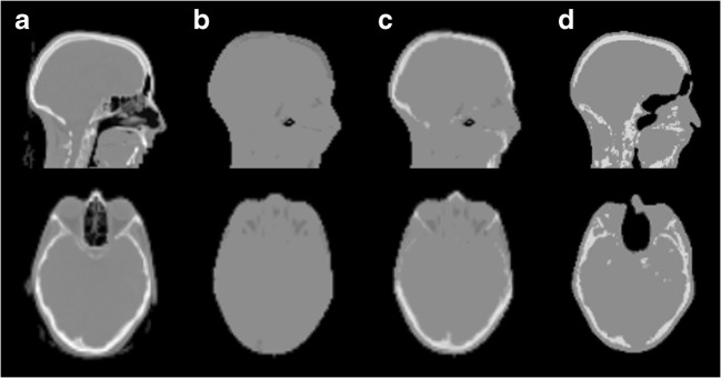
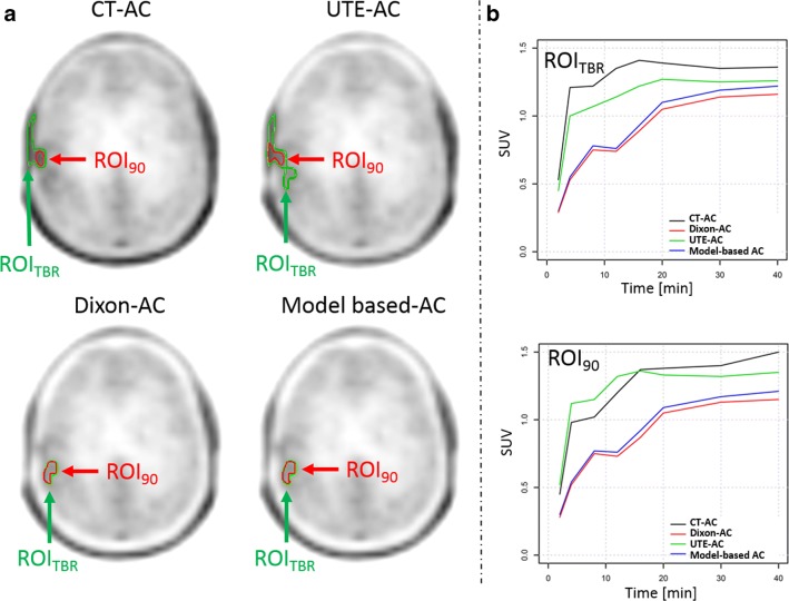
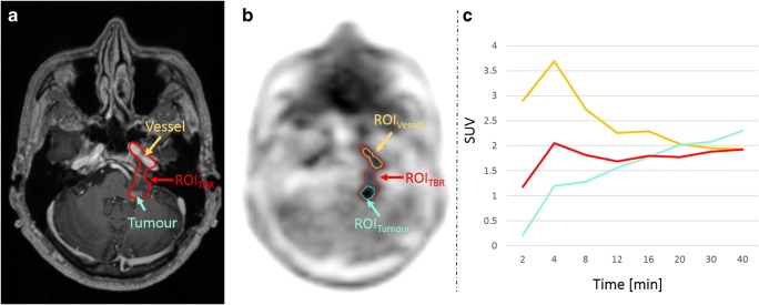
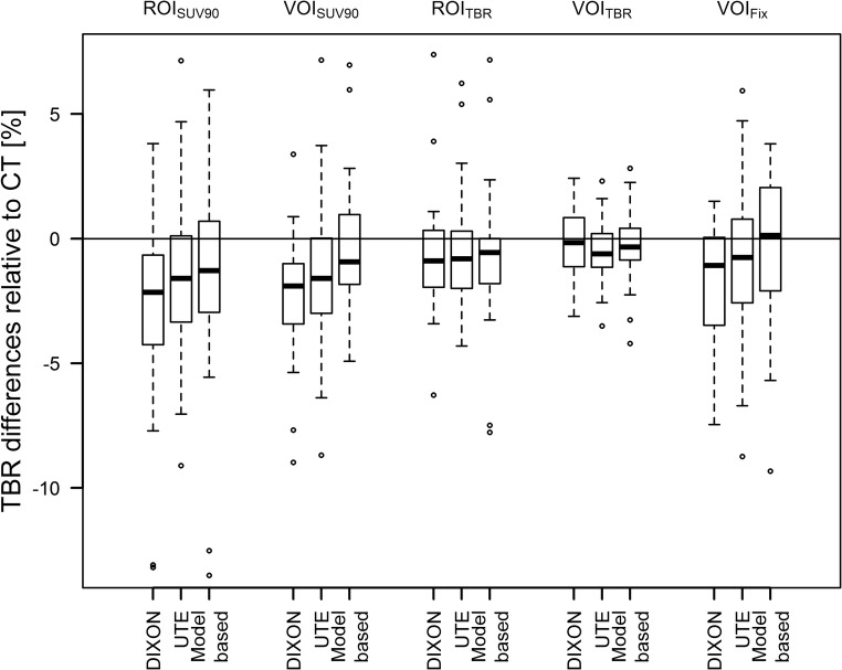
Twenty-four patients with suspected brain tumours underwent dynamic [18F]FET-PET/MRI examinations and subsequent low-dose computed tomography (CT) scans of the head. The dynamic PET data was reconstructed using the following AC methods: standard Dixon-based AC and ultra-short echo time MRI-based AC (MR-AC) and a model-based AC approach. All data were reconstructed also using CT-based AC (reference). For all lesions and reconstructions, time-activity curves (TACs) and time to peak (TTP) were extracted using different region-of-interest (ROI) and volume-of-interest (VOI) definitions. According to the most common evaluation approaches, TACs were categorised into two or three distinct curve patterns. Changes in TTP and TAC patterns compared to PET using CT-based AC were reported.
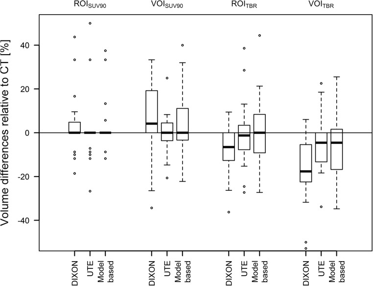
In the majority of cases, TAC patterns did not change. However, TAC pattern changes as well as changes in TTP were observed in up to 8% and 17% of the cases when using different MR-AC methods and ROI/VOI definitions, respectively. However, these changes in TTP and TAC pattern were attributed to different delineations of the ROIs/VOIs in PET corrected with different AC methods.
PET/MRI using different MR-AC methods can be used for the assessment of TAC patterns in dynamic [18F]FET studies, as long as a meaningful delineation of the area of interest within the tumour is ensured.
• PET/MRI using different MR-AC methods can be used for dynamic [18F]FET studies. • A meaningful segmentation of the area of interest needs to be ensured, mandating a visual validation of the delineation by an experienced reader.
评估基于动态 [18F]FET 正电子发射断层扫描/磁共振成像(PET/MRI)研究的肿瘤分级是否受不同基于 MRI 的衰减校正(AC)方法的影响。
24 例疑似脑肿瘤患者接受了动态 [18F]FET-PET/MRI 检查和随后的头部低剂量 CT(CT)扫描。动态 PET 数据使用以下 AC 方法进行重建:标准 Dixon 基于 AC 和超短回波时间 MRI 基于 AC(MR-AC)和基于模型的 AC 方法。所有数据均使用 CT 基于 AC(参考)进行重建。对于所有病变和重建,使用不同的感兴趣区域(ROI)和感兴趣体积(VOI)定义提取时间活性曲线(TAC)和达峰时间(TTP)。根据最常见的评估方法,将 TAC 分为两种或三种不同的曲线模式。报告了与使用 CT 基于 AC 的 PET 相比 TTP 和 TAC 模式的变化。
在大多数情况下,TAC 模式没有改变。然而,当使用不同的 MR-AC 方法和 ROI/VOI 定义时,高达 8%和 17%的病例观察到 TAC 模式变化和 TTP 变化。然而,这些 TTP 和 TAC 模式的变化归因于使用不同 AC 方法校正的 PET 中 ROI/VOI 的不同描绘。
使用不同的 MR-AC 方法的 PET/MRI 可用于评估动态 [18F]FET 研究中的 TAC 模式,只要确保在肿瘤内有意义地描绘感兴趣区域。
• 使用不同的 MR-AC 方法的 PET/MRI 可用于动态 [18F]FET 研究。• 需要确保有意义地描绘感兴趣区域,这需要由有经验的读者进行视觉验证。