• 文献检索
  • 文档翻译
  • 深度研究
  • 学术资讯
  • Suppr Zotero 插件Zotero 插件
  • 邀请有礼
  • 套餐&价格
  • 历史记录
应用&插件
Suppr Zotero 插件Zotero 插件浏览器插件Mac 客户端Windows 客户端微信小程序
定价
高级版会员购买积分包购买API积分包
服务
文献检索文档翻译深度研究API 文档MCP 服务
关于我们
关于 Suppr公司介绍联系我们用户协议隐私条款
关注我们

Suppr 超能文献

核心技术专利:CN118964589B侵权必究
粤ICP备2023148730 号-1Suppr @ 2026

文献检索

告别复杂PubMed语法,用中文像聊天一样搜索,搜遍4000万医学文献。AI智能推荐,让科研检索更轻松。

立即免费搜索

文件翻译

保留排版,准确专业,支持PDF/Word/PPT等文件格式,支持 12+语言互译。

免费翻译文档

深度研究

AI帮你快速写综述,25分钟生成高质量综述,智能提取关键信息,辅助科研写作。

立即免费体验

相似文献

1
Rapid loss of perihematomal cell viability in the collagenase intracerebral hemorrhage model.胶原酶脑出血模型中血肿周围细胞活力迅速丧失。
Brain Res. 2019 May 15;1711:91-96. doi: 10.1016/j.brainres.2019.01.014. Epub 2019 Jan 10.
2
Systemic hemin therapy attenuates blood-brain barrier disruption after intracerebral hemorrhage.全身血红素疗法可减轻脑出血后血脑屏障的破坏。
Neurobiol Dis. 2014 Oct;70:245-51. doi: 10.1016/j.nbd.2014.06.005. Epub 2014 Jun 18.
3
Astrocyte heme oxygenase-1 reduces mortality and improves outcome after collagenase-induced intracerebral hemorrhage.星形胶质细胞血红素加氧酶-1可降低胶原酶诱导的脑出血后的死亡率并改善预后。
Neurobiol Dis. 2017 Jun;102:140-146. doi: 10.1016/j.nbd.2017.03.008. Epub 2017 Mar 18.
4
Modeling intracerebral hemorrhage in mice: injection of autologous blood or bacterial collagenase.小鼠脑出血建模:自体血注射或细菌胶原酶注射。
J Vis Exp. 2012 Sep 22(67):e4289. doi: 10.3791/4289.
5
Effect of hemopexin treatment on outcome after intracerebral hemorrhage in mice.血红素结合蛋白治疗对小鼠脑出血后结局的影响。
Brain Res. 2021 Aug 15;1765:147507. doi: 10.1016/j.brainres.2021.147507. Epub 2021 Apr 28.
6
Celecoxib induces functional recovery after intracerebral hemorrhage with reduction of brain edema and perihematomal cell death.塞来昔布可减轻脑水肿和血肿周围细胞死亡,从而促进脑出血后的功能恢复。
J Cereb Blood Flow Metab. 2004 Aug;24(8):926-33. doi: 10.1097/01.WCB.0000130866.25040.7D.
7
Increased striatal injury and behavioral deficits after intracerebral hemorrhage in hemopexin knockout mice.载脂蛋白缺失小鼠脑出血后纹状体损伤增加和行为缺陷。
J Neurosurg. 2011 Apr;114(4):1159-67. doi: 10.3171/2010.10.JNS10861. Epub 2010 Dec 3.
8
Astrocyte overexpression of heme oxygenase-1 improves outcome after intracerebral hemorrhage.星形胶质细胞中血红素加氧酶-1的过表达可改善脑出血后的预后。
Stroke. 2015 Apr;46(4):1093-8. doi: 10.1161/STROKEAHA.115.008686. Epub 2015 Feb 17.
9
Iron regulatory protein-2 knockout increases perihematomal ferritin expression and cell viability after intracerebral hemorrhage.铁调节蛋白-2 敲除增加脑出血后血肿周围铁蛋白表达和细胞活力。
Brain Res. 2010 Jun 14;1337:95-103. doi: 10.1016/j.brainres.2010.04.018. Epub 2010 Apr 24.
10
Failure of deferoxamine, an iron chelator, to improve outcome after collagenase-induced intracerebral hemorrhage in rats.铁螯合剂去铁胺治疗胶原酶诱导的大鼠脑出血失败。
Brain Res. 2010 Jan 14;1309:95-103. doi: 10.1016/j.brainres.2009.10.058. Epub 2009 Oct 30.

引用本文的文献

1
Long Noncoding RNA GAS5-Involved Progression of Neonatal Hydrocephalus and Inflammatory Responses.长链非编码RNA GAS5参与新生儿脑积水进展及炎症反应
Mol Biotechnol. 2025 Feb;67(2):661-672. doi: 10.1007/s12033-024-01077-x. Epub 2024 Mar 1.
2
Effect of hemopexin treatment on outcome after intracerebral hemorrhage in mice.血红素结合蛋白治疗对小鼠脑出血后结局的影响。
Brain Res. 2021 Aug 15;1765:147507. doi: 10.1016/j.brainres.2021.147507. Epub 2021 Apr 28.
3
Annexin A2 depletion exacerbates the intracerebral microhemorrhage induced by acute rickettsia and Ebola virus infections.膜联蛋白 A2 耗竭加剧急性立克次体和埃博拉病毒感染引起的脑内微出血。
PLoS Negl Trop Dis. 2020 Jul 20;14(7):e0007960. doi: 10.1371/journal.pntd.0007960. eCollection 2020 Jul.

本文引用的文献

1
Intracerebral Hemorrhage in Mice.小鼠脑出血
Methods Mol Biol. 2018;1717:83-91. doi: 10.1007/978-1-4939-7526-6_7.
2
Inhibition of astrocytic activity alleviates sequela in acute stages of intracerebral hemorrhage.抑制星形胶质细胞活性可减轻脑出血急性期的后遗症。
Oncotarget. 2017 Oct 24;8(55):94850-94861. doi: 10.18632/oncotarget.22022. eCollection 2017 Nov 7.
3
Astrocyte heme oxygenase-1 reduces mortality and improves outcome after collagenase-induced intracerebral hemorrhage.星形胶质细胞血红素加氧酶-1可降低胶原酶诱导的脑出血后的死亡率并改善预后。
Neurobiol Dis. 2017 Jun;102:140-146. doi: 10.1016/j.nbd.2017.03.008. Epub 2017 Mar 18.
4
Targeting the Nrf2-Heme Oxygenase-1 Axis after Intracerebral Hemorrhage.脑出血后靶向Nrf2-血红素加氧酶-1轴
Curr Pharm Des. 2017;23(15):2226-2237. doi: 10.2174/1381612822666161027150616.
5
Efficacy of deferoxamine in animal models of intracerebral hemorrhage: a systematic review and stratified meta-analysis.去铁胺在脑出血动物模型中的疗效:一项系统评价与分层荟萃分析
PLoS One. 2015 May 22;10(5):e0127256. doi: 10.1371/journal.pone.0127256. eCollection 2015.
6
Astrocyte overexpression of heme oxygenase-1 improves outcome after intracerebral hemorrhage.星形胶质细胞中血红素加氧酶-1的过表达可改善脑出血后的预后。
Stroke. 2015 Apr;46(4):1093-8. doi: 10.1161/STROKEAHA.115.008686. Epub 2015 Feb 17.
7
Sex differences in incidence, pathophysiology, and outcome of primary intracerebral hemorrhage.原发性脑出血在发病率、病理生理学及预后方面的性别差异。
Stroke. 2015 Mar;46(3):886-92. doi: 10.1161/STROKEAHA.114.007682. Epub 2015 Feb 5.
8
Neuroprotective effect of heme oxygenase-2 knockout in the blood injection model of intracerebral hemorrhage.脑出血血液注射模型中血红素加氧酶-2基因敲除的神经保护作用
BMC Res Notes. 2014 Aug 22;7:561. doi: 10.1186/1756-0500-7-561.
9
Systemic hemin therapy attenuates blood-brain barrier disruption after intracerebral hemorrhage.全身血红素疗法可减轻脑出血后血脑屏障的破坏。
Neurobiol Dis. 2014 Oct;70:245-51. doi: 10.1016/j.nbd.2014.06.005. Epub 2014 Jun 18.
10
Prostaglandin F2α FP receptor antagonist improves outcomes after experimental traumatic brain injury.前列腺素 F2α FP 受体拮抗剂可改善实验性创伤性脑损伤的预后。
J Neuroinflammation. 2013 Oct 30;10:132. doi: 10.1186/1742-2094-10-132.

胶原酶脑出血模型中血肿周围细胞活力迅速丧失。

Rapid loss of perihematomal cell viability in the collagenase intracerebral hemorrhage model.

机构信息

Department of Emergency Medicine, Thomas Jefferson University, USA.

Department of Emergency Medicine, Thomas Jefferson University, USA.

出版信息

Brain Res. 2019 May 15;1711:91-96. doi: 10.1016/j.brainres.2019.01.014. Epub 2019 Jan 10.

DOI:10.1016/j.brainres.2019.01.014
PMID:30639124
原文链接:https://pmc.ncbi.nlm.nih.gov/articles/PMC6519080/
Abstract

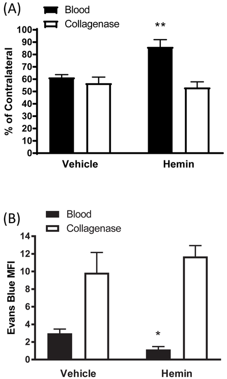
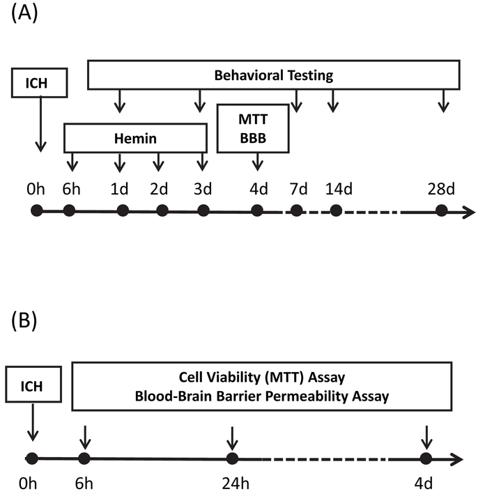
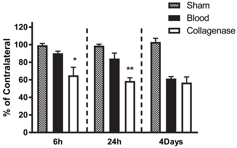
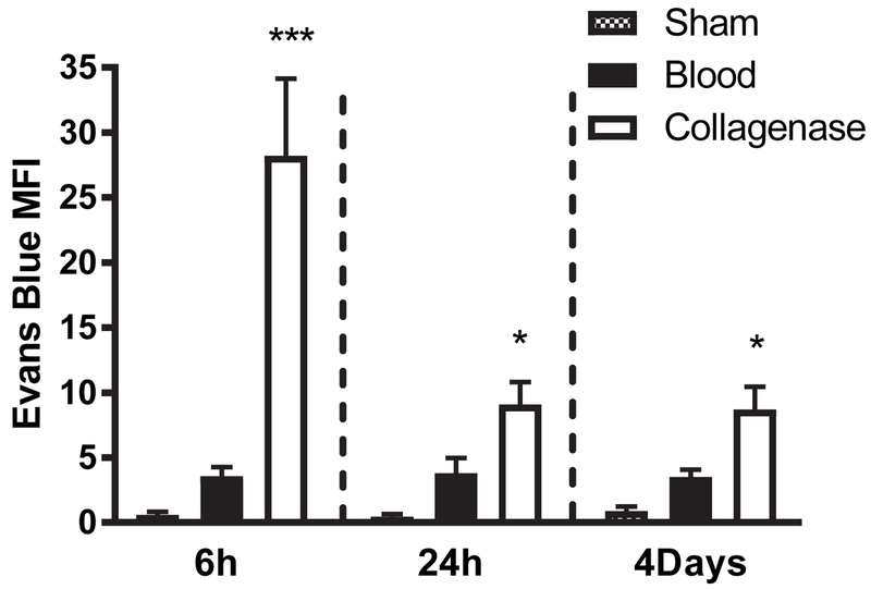
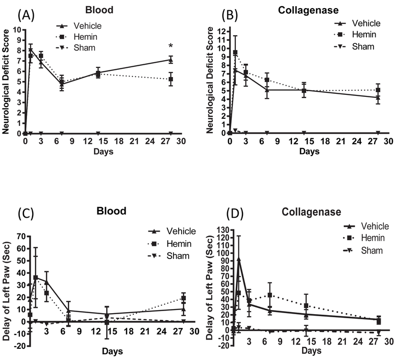
The effective time window of any therapeutic in an experimental stroke model is limited by the rate of injury progression. Intracerebral hemorrhage in rodents is commonly induced by striatal injection of either autologous blood or bacterial collagenase, which digests local blood vessels. During time window studies of the heme oxygenase-1 inducer hemin, which is protective when administered within 1-3 h in both models, the rate of perihematomal injury was directly compared after striatal blood or collagenase injection. Surprisingly, about 80% of the loss of perihematomal cell viability as measured by MTT reduction assay occurred within 6 h of collagenase injection. In contrast, significant viability loss was not observed at this time point after autologous blood injection, but rather it progressed over the subsequent four days to a level similar to that produced by collagenase. Consistent with these observations, systemic hemin therapy reduced blood-brain barrier disruption and perihematomal cell injury when initiated at 6 h after striatal injection of blood but not collagenase. These results indicate that the rate of early cell injury differs markedly in the collagenase and blood injection ICH models, which may contribute to inconsistent results in time window studies. The blood injection model may be more appropriate for prolonged time window studies of a neuroprotective agent.

摘要

任何治疗方法在实验性中风模型中的有效时间窗口都受到损伤进展速度的限制。在啮齿动物中,脑内出血通常通过纹状体注射自体血液或细菌胶原酶来诱导,后者可消化局部血管。在血红素加氧酶-1诱导剂血红素的时间窗研究中,该药物在两种模型中均在 1-3 小时内给药具有保护作用,在纹状体血液或胶原酶注射后直接比较了血肿周围损伤的速度。令人惊讶的是,用 MTT 还原测定法测量的血肿周围细胞活力丧失约 80%发生在胶原酶注射后 6 小时内。相比之下,在这个时间点,自体血注射后没有观察到明显的细胞活力丧失,而是在随后的四天内逐渐增加到与胶原酶相似的水平。与这些观察结果一致,全身性血红素治疗在纹状体注射血液后 6 小时开始时可降低血脑屏障破坏和血肿周围细胞损伤,但在注射胶原酶时则无效。这些结果表明,胶原酶和血液注射 ICH 模型中的早期细胞损伤速度差异显著,这可能导致时间窗研究中的结果不一致。血液注射模型可能更适合于神经保护剂的长时间窗研究。

https://cdn.ncbi.nlm.nih.gov/pmc/blobs/698b/6519080/9e1df2141f91/nihms-1519479-f0005.jpg
https://cdn.ncbi.nlm.nih.gov/pmc/blobs/698b/6519080/1dada229c156/nihms-1519479-f0001.jpg
https://cdn.ncbi.nlm.nih.gov/pmc/blobs/698b/6519080/58eaf8334c0c/nihms-1519479-f0002.jpg
https://cdn.ncbi.nlm.nih.gov/pmc/blobs/698b/6519080/3dec519dcf9b/nihms-1519479-f0003.jpg
https://cdn.ncbi.nlm.nih.gov/pmc/blobs/698b/6519080/820ec3a63543/nihms-1519479-f0004.jpg
https://cdn.ncbi.nlm.nih.gov/pmc/blobs/698b/6519080/9e1df2141f91/nihms-1519479-f0005.jpg