Department of Pediatrics, University of Wisconsin School of Medicine and Public Health, Madison, WI, USA; Waisman Center, University of Wisconsin School of Medicine and Public Health, Madison, WI, USA.
Waisman Center, University of Wisconsin School of Medicine and Public Health, Madison, WI, USA.
Neurochem Int. 2019 Jul;127:137-147. doi: 10.1016/j.neuint.2018.12.016. Epub 2019 Jan 9.
Neuroinflammation plays an important role in ischemic brain injury and recovery, however the interplay between brain development and the neuroinflammatory response is poorly understood. We previously described age-dependent differences in the microglial response and the effect of microglial inhibition. Here we investigate whether age-dependent microglial responses may be related to pre-injury developmental differences in microglial phenotype.
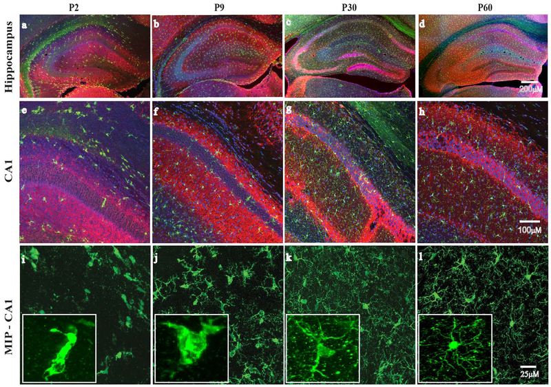
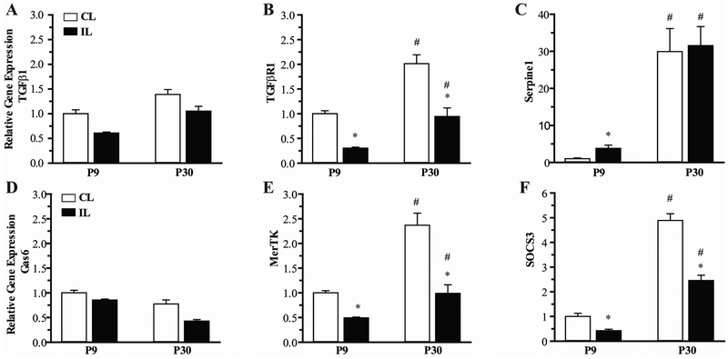
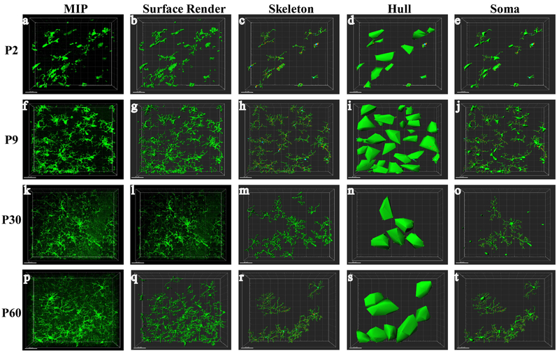
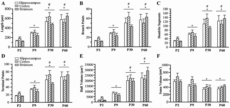
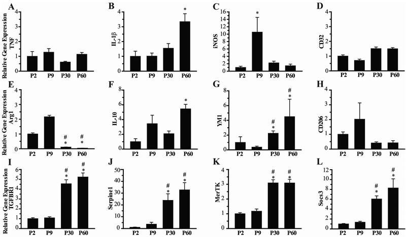
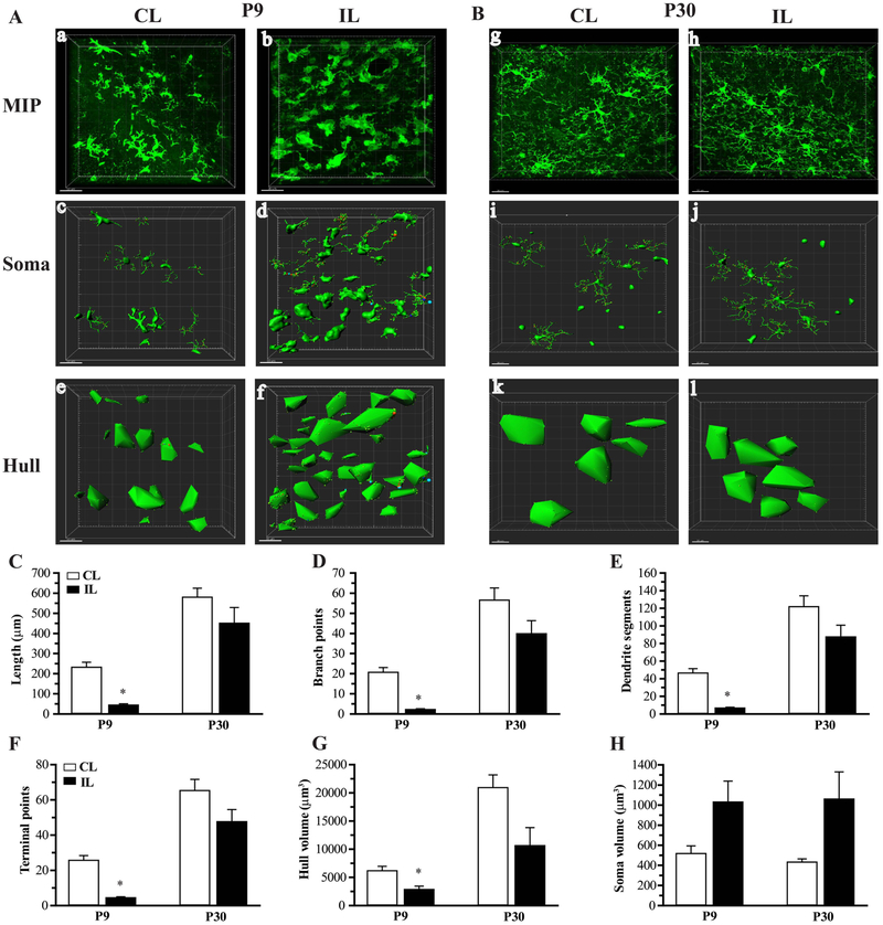
Measures of microglia morphology were quantified using semi-automated software analysis of immunostained sections from postnatal day 2 (P2), P9, P30 and P60 mice using IMARIS. Microglia were isolated from P2, P9, P30 and P60 mice, and expression of markers of classical and alternative microglial activation was assessed, as well as transforming growth factor beta (TGF-β) receptor, Serpine1, Mer Tyrosine Kinase (MerTK), and the suppressor of cytokine signaling (SOCS3). Hypoxia-ischemia (HI) was induced in P9 and P30 mice using unilateral carotid artery ligation and exposure to 10% oxygen for 50 min. Microglia morphology and microglial expression of genes in the TGF-β and MerTK pathways were determined in ipsilateral and contralateral hippocampus.
A progressive and significant increase in microglia branching morphology was seen in all brain regions from P2 to P30. No consistent classical or alternative activation profile was seen in isolated microglia. A clear transition to increased expression of TGF-β and its downstream effector serpine1 was seen between P9 and P30. A similar increase in expression was seen in MerTK and its downstream effector SOCS3. HI resulted in a significant decrease in branching morphology only in the P9 mice, and expression of TGF-β receptor, Serpine1, MerTK, and SOCS3 were elevated in P30 mice compared to P9 post-HI.
Microglia maturation is associated with changes in morphology and gene expression, and microglial responses to ischemia in the developing brain differ based on the age at which injury occurs.
神经炎症在缺血性脑损伤和恢复中起着重要作用,然而,大脑发育和神经炎症反应之间的相互作用还知之甚少。我们之前描述了小胶质细胞反应的年龄依赖性差异以及小胶质细胞抑制的作用。在这里,我们研究年龄依赖性小胶质细胞反应是否与损伤前发育过程中小胶质细胞表型的差异有关。
使用 IMARIS 对来自出生后第 2 天(P2)、P9、P30 和 P60 天的小鼠免疫染色切片进行半自动软件分析,对小胶质细胞形态进行定量测量。从小鼠 P2、P9、P30 和 P60 中分离小胶质细胞,评估经典和替代小胶质细胞激活的标志物,以及转化生长因子-β(TGF-β)受体、Serpine1、Mer 酪氨酸激酶(MerTK)和细胞因子信号转导抑制因子(SOCS3)。在 P9 和 P30 天的小鼠中,通过单侧颈总动脉结扎和暴露于 10%氧气 50 分钟来诱导缺氧缺血(HI)。在同侧和对侧海马体中确定 HI 后小胶质细胞形态和 TGF-β 和 MerTK 通路中的基因表达。
从小鼠 P2 到 P30 所有脑区的小胶质细胞分支形态均呈进行性和显著增加。从 P2 到 P30 未观察到一致的经典或替代激活特征。在 P9 和 P30 之间观察到 TGF-β及其下游效应物 Serpine1 的表达明显增加。在 MerTK 及其下游效应物 SOCS3 中也观察到类似的增加。仅在 P9 天的小鼠中观察到分支形态明显减少,与 P9 天相比,P30 天的 HI 小鼠 TGF-β 受体、Serpine1、MerTK 和 SOCS3 的表达增加。
小胶质细胞成熟与形态和基因表达的变化有关,并且发育中的大脑对缺血的小胶质细胞反应因损伤发生的年龄而异。