Department of Histology and Embryology, Pomeranian Medical University, Szczecin, Poland.
Department of Biology and Medical Parasitology, Pomeranian Medical University, Szczecin, Poland.
Biol Trace Elem Res. 2019 Oct;191(2):300-305. doi: 10.1007/s12011-018-1621-6. Epub 2019 Jan 15.
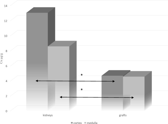
In the body, disorders in the composition and concentration of trace elements, including copper, can lead to the development of various alterations that may result in incorrect functioning of the kidneys. Data on the concentrations of copper in human kidneys are discussed; however, little is known about the concentration of trace elements within rejected renal grafts and kidneys with tumor lesions. The aim of our study was to compare the copper concentration between cancerous kidneys and rejected renal grafts with the division on renal cortex and renal medulla. Material consisted of kidneys from patients hospitalized at the Department of Urology and General Surgery and Transplantation of the Independent Public Clinical Hospital No. 2 at the Pomeranian Medical University in Szczecin, north-western Poland. The study material consisted of kidneys with tumor lesions (n = 33), and renal grafts (n = 10), obtained from patients belongs to the north-western areas of Poland. The examination was performed using ICP-AES method. Regarding the pathological kidneys, excluding grafts, the concentration of Cu in the renal cortex was 52% higher than in medullary region and the difference between the compared concentrations was statistically confirmed (p < 0.05). Taking into account renal grafts, the concentration of Cu in the medulla was slightly lower than in the cortex (less than 3%). In summary, copper in rejected and cancerous kidneys tends to accumulate in higher amount in the renal cortex than medulla, what can be explained by the fact that renal corpuscles, where the first phase of filtration is performed, are located only in the cortical region of the kidney. Furthermore, renal grafts accumulate significantly less copper than kidneys with neoplastic changes, what could have been caused by immunosuppressive medicines used by the graft recipients. The lower copper concentration in renal grafts could be a consequence of the altered immune system, including inflammatory process or/and non-immune mechanisms. Additionally, cancerous and non-cancerous kidneys exhibit different perfusion rate in renal glomeruli, what can finally lead to disparity in chemical elements concentration, including copper.
在体内,包括铜在内的微量元素的组成和浓度的紊乱会导致各种变化的发生,这些变化可能导致肾脏功能不正常。本文讨论了人类肾脏中铜的浓度,但对于肿瘤病变肾脏和被排斥的移植肾脏中微量元素的浓度知之甚少。我们的研究目的是比较癌症肾脏和被排斥的移植肾脏中铜的浓度,将其分为肾皮质和肾髓质两部分。研究材料包括来自波兰西北部什切青独立公共临床医院第 2 泌尿科和普通外科及移植科住院患者的肾脏。研究材料包括 33 例肿瘤病变肾脏和 10 例肾移植,这些患者来自波兰西北部地区。使用 ICP-AES 法进行检查。对于非移植的病理性肾脏,Cu 在肾皮质中的浓度比在髓质中高 52%,且比较浓度之间的差异具有统计学意义(p<0.05)。考虑到肾移植,Cu 在髓质中的浓度略低于皮质(不到 3%)。总之,在被排斥和癌变的肾脏中,铜在肾皮质中的积累量比在髓质中多,这可以用发生在肾小球中的第一阶段过滤仅位于肾脏皮质区域的事实来解释。此外,移植肾积累的铜明显少于发生肿瘤变化的肾脏,这可能是由于移植受者使用免疫抑制药物所致。移植肾中铜浓度较低可能是免疫系统改变的结果,包括炎症过程或/和非免疫机制。此外,癌症和非癌症肾脏的肾小球灌注率不同,这最终可能导致包括铜在内的化学元素浓度的差异。