Department of Neurological Surgery, Emory University School of Medicine, Atlanta, Georgia.
Department of Neurology, Emory University School of Medicine, Atlanta, Georgia.
Epilepsia. 2019 Feb;60(2):220-232. doi: 10.1111/epi.14634. Epub 2019 Jan 17.
Magnetic resonance (MR) thermography-guided laser interstitial thermal therapy, or stereotactic laser ablation (SLA), is a minimally invasive alternative to open surgery for focal epilepsy caused by cerebral cavernous malformations (CCMs). We examined the safety and effectiveness of SLA of epileptogenic CCMs.
We retrospectively analyzed 19 consecutive patients who presented with focal seizures associated with a CCM. Each patient underwent SLA of the CCM and adjacent cortex followed by standard clinical and imaging follow-up.
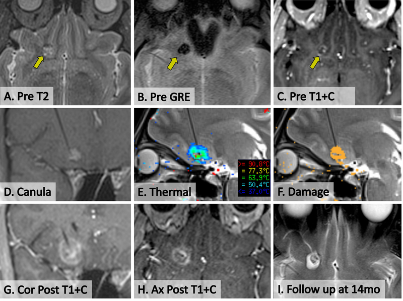
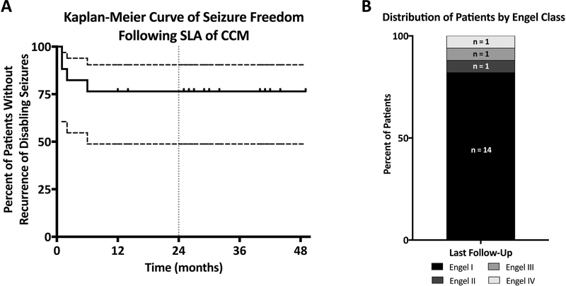
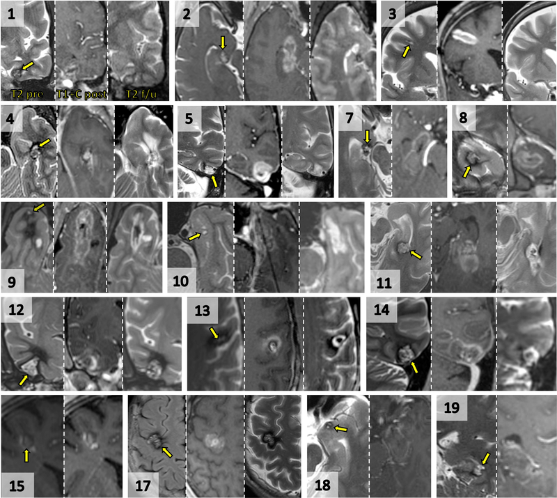
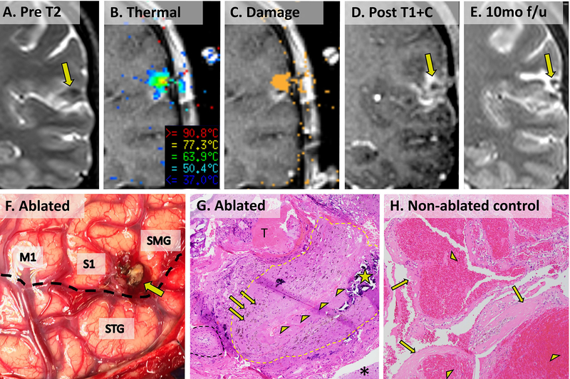
All but one patient had chronic medically refractory epilepsy (median duration 8 years, range 0.5-52 years). Lesions were located in the temporal (13), frontal (five), and parietal (one) lobes. CCMs induced magnetic susceptibility artifacts during thermometry, but perilesional cortex was easily visualized. Fourteen of 17 patients (82%) with >12 months of follow-up achieved Engel class I outcomes, of which 10 (59%) were Engel class IA. Two patients who were not seizure-free from SLA alone became so following intracranial electrode-guided open resection. Delayed postsurgical imaging validated CCM involution (median 83% volume reduction) and ablation of surrounding cortex. Histopathologic examination of one previously ablated CCM following open surgery confirmed obliteration. SLA caused no detectable hemorrhages. Two symptomatic neurologic deficits (visual and motor) were predictable, and neither was permanently disabling.
In a consecutive retrospective series, MR thermography-guided SLA was an effective alternative to open surgery for epileptogenic CCM. The approach was free of hemorrhagic complications, and clinically significant neurologic deficits were predictable. SLA presents no barrier to subsequent open surgery when needed.
磁共振(MR)热成像引导激光间质热疗或立体定向激光消融(SLA)是一种针对由脑海绵状血管畸形(CCM)引起的局灶性癫痫的微创替代开颅手术方法。我们研究了 SLA 治疗致痫性 CCM 的安全性和有效性。
我们回顾性分析了 19 例因 CCM 而出现局灶性癫痫发作的连续患者。每位患者均接受了 CCM 和相邻皮质的 SLA,随后进行了标准的临床和影像学随访。
除 1 例患者外,其余患者均患有慢性药物难治性癫痫(中位病程 8 年,范围 0.5-52 年)。病变位于颞叶(13 例)、额叶(5 例)和顶叶(1 例)。CCM 在测温过程中会产生磁敏感性伪影,但周边皮质很容易被看到。17 例中有 14 例(82%)随访时间>12 个月的患者达到了 Engel Ⅰ级结果,其中 10 例(59%)为 Engel ⅠA 级。2 例单纯 SLA 术后仍有癫痫发作的患者在接受颅内电极引导下的开颅切除术后也无癫痫发作。术后延迟影像学检查证实 CCM 消退(中位体积减少 83%)和周边皮质消融。对 1 例先前经开颅手术切除的 CCM 进行组织病理学检查证实其已闭塞。SLA 未引起可检测到的出血。2 例有症状的神经功能缺损(视觉和运动)是可预测的,且均无永久性残疾。
在连续的回顾性系列研究中,MR 热成像引导的 SLA 是治疗致痫性 CCM 的一种有效替代开颅手术的方法。该方法无出血并发症,且可预测到有临床意义的神经功能缺损。当需要时,SLA 不会对后续的开颅手术造成障碍。