Fan Bo, Shi Sifeng, Shen Xiaofei, Yang Xiaolong, Liu Na, Wu Guixia, Guo Xiaojuan, Huang Ning
Department of Pathophysiology, Research Unit of Infection and Immunity, West China College of Basic and Forensic Medicine, Sichuan University, Chengdu, Sichuan 610041, P.R. China.
Department of Pathophysiology, Xuzhou Medical University, Xuzhou, Jiangsu 221000, P.R. China.
Oncol Lett. 2019 Jan;17(1):1160-1166. doi: 10.3892/ol.2018.9668. Epub 2018 Nov 5.
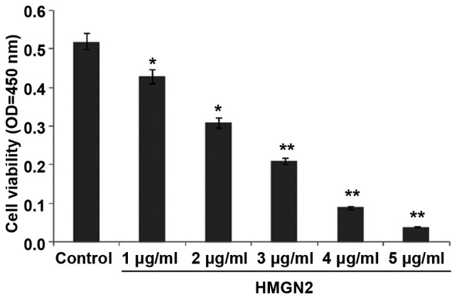
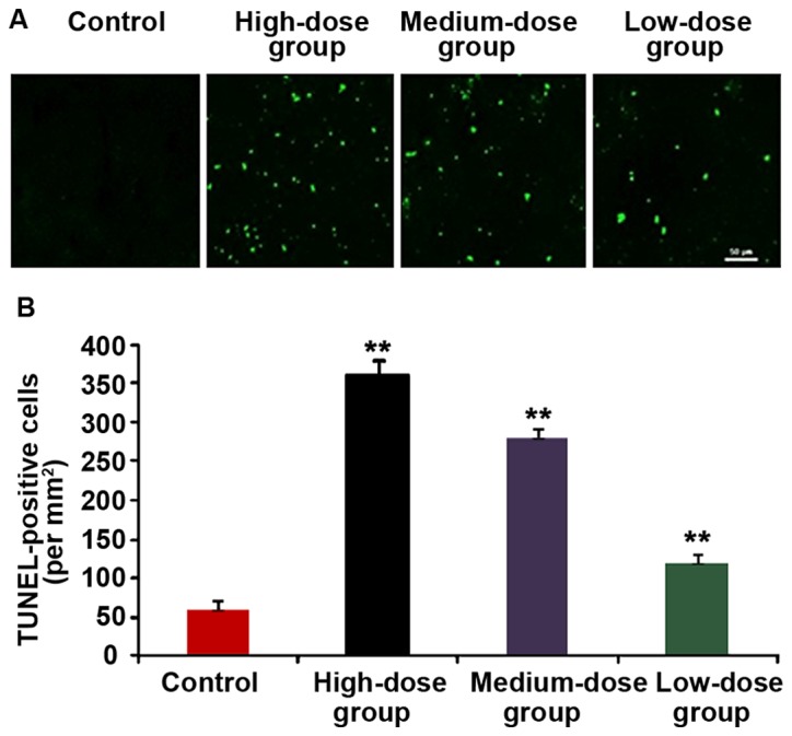
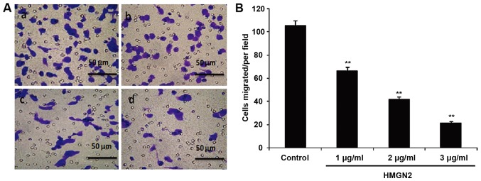
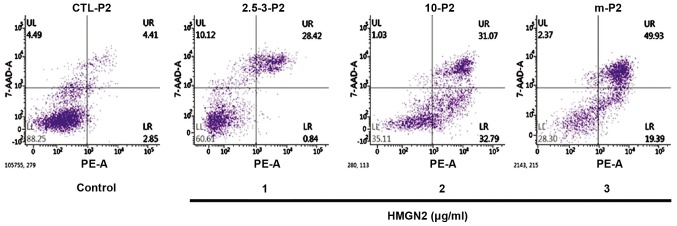
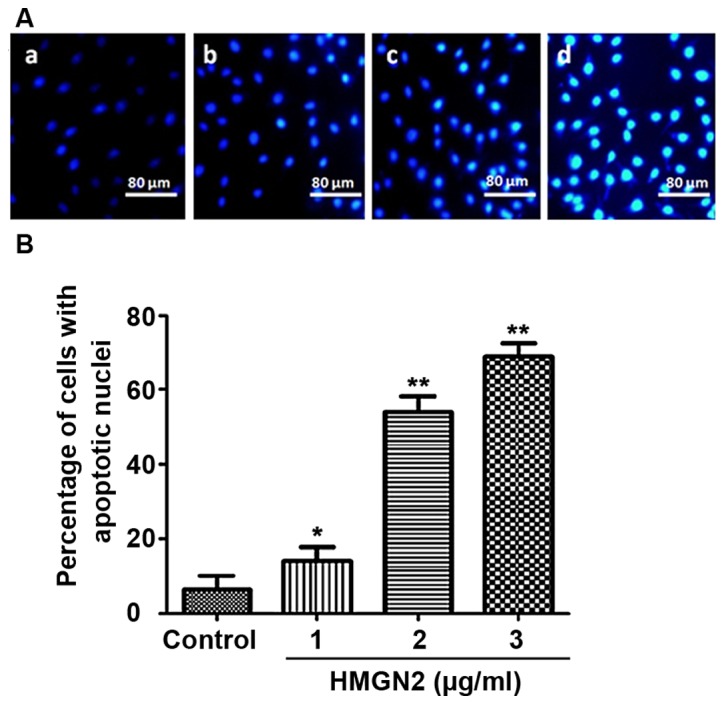
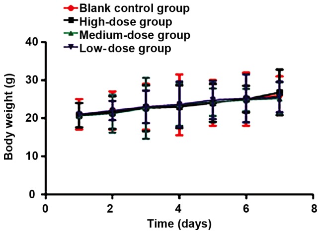
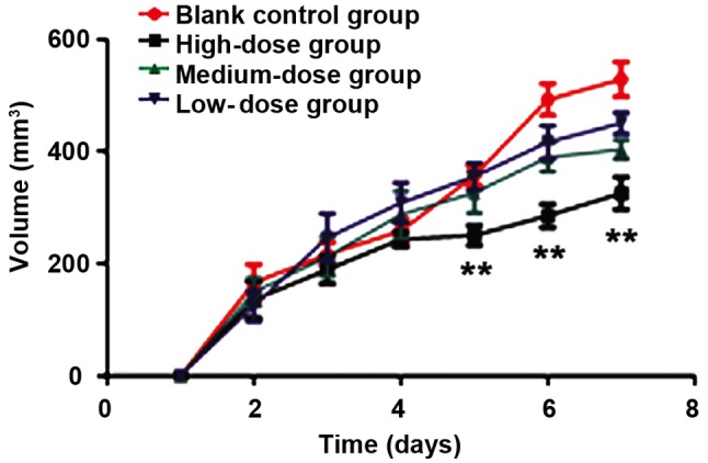
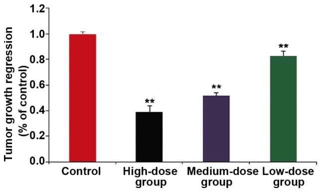
We investigated the effect of high mobility group protein N2 (HMGN2) on the proliferation and apoptosis of the human MCF-7 breast cancer cell line, and its effect on tumor growth in a subcutaneous heterotopic transplantation tumor model of breast cancer. The cell viability assay was used to verify the effect of the recombinant human HMGN2 on MCF-7 cell proliferation. The Transwell chamber assay was used to verify the effect of HMGN2 on MCF-7 cell migration. Flow cytometry and Hoechst staining were used to detect the effect of HMGN2 on MCF-7 cell apoptosis. MCF-7 was injected to establish a subcutaneous heterotopic transplantation tumor model of breast cancer in nude mice. The size, weight and volume of tumor in each group were compared after the administration of different concentrations of HMGN2 solution around the tumor tissue at day 1, 3, 5 and 7. The tumor tissue was removed and cut into sections, and the apoptotic cells in tumors of nude mice were detected by a TUNEL kit. The CCK-8 assay showed that HMGN2 at different concentrations inhibited the proliferation of the MCF-7 breast cancer cells, and the proliferation of MCF-7 cells were significantly inhibited when the concentration of HMGN2 reached 3 µg/ml (P<0.01). The Transwell chamber assay showed that 3 µg/ml of HMGN2 significantly decreased the migration capacity of MCF-7 cells (P<0.01). Flow cytometry and Hoechst staining showed that 3 µg/ml of HMGN2 significantly increased apoptosis of MCF-7 cells (P<0.01). After the nude mouse model of breast cancer was established, HMGN2 at different concentrations was injected around the tumor tissue at day 1, 3, 5 and 7. We demonstrated that the growth of breast cancer was significantly inhibited when the concentration of HMGN2 reached 15 µg/ml. TUNEL staining showed that the number of apoptotic cells in the 15 µg/ml dose group was significantly higher than that in the control group (P<0.01). Therefore, and experiments proved that recombinant human HMGN2 could significantly inhibit the proliferation and migration of breast cancer cells, which increased the apoptosis of breast cancer cells and exerted anti-breast cancer effects, which enriched our understanding of the biological roles of HMGN2.
我们研究了高迁移率族蛋白N2(HMGN2)对人MCF-7乳腺癌细胞系增殖和凋亡的影响,及其在乳腺癌皮下异位移植瘤模型中对肿瘤生长的影响。采用细胞活力测定法验证重组人HMGN2对MCF-7细胞增殖的影响。采用Transwell小室测定法验证HMGN2对MCF-7细胞迁移的影响。采用流式细胞术和Hoechst染色检测HMGN2对MCF-7细胞凋亡的影响。将MCF-7注射到裸鼠体内建立乳腺癌皮下异位移植瘤模型。在第1、3、5和7天,在肿瘤组织周围给予不同浓度的HMGN2溶液后,比较各组肿瘤的大小、重量和体积。取出肿瘤组织并切成切片,用TUNEL试剂盒检测裸鼠肿瘤中的凋亡细胞。CCK-8测定结果显示,不同浓度的HMGN2均抑制MCF-7乳腺癌细胞的增殖,当HMGN2浓度达到3μg/ml时,MCF-7细胞的增殖受到显著抑制(P<0.01)。Transwell小室测定结果显示,3μg/ml的HMGN2显著降低MCF-7细胞的迁移能力(P<0.01)。流式细胞术和Hoechst染色显示,3μg/ml的HMGN2显著增加MCF-7细胞的凋亡(P<0.01)。建立乳腺癌裸鼠模型后,在第1、3、5和7天在肿瘤组织周围注射不同浓度的HMGN2。我们证明,当HMGN2浓度达到15μg/ml时,乳腺癌的生长受到显著抑制。TUNEL染色显示,15μg/ml剂量组的凋亡细胞数量显著高于对照组(P<¬0.01)。因此,体内和体外实验证明,重组人HMGN2可显著抑制乳腺癌细胞的增殖和迁移,增加乳腺癌细胞的凋亡,发挥抗乳腺癌作用,这丰富了我们对HMGN2生物学作用的认识。