Urology Department, Yale University School of Medicine, New Haven, CT.
Department of Pathology & Laboratory Medicine, The Warren Alpert Medical School of Brown University and The Miriam Hospital, Providence, RI.
Urology. 2020 May;139:134-140. doi: 10.1016/j.urology.2019.01.008. Epub 2019 Jan 16.
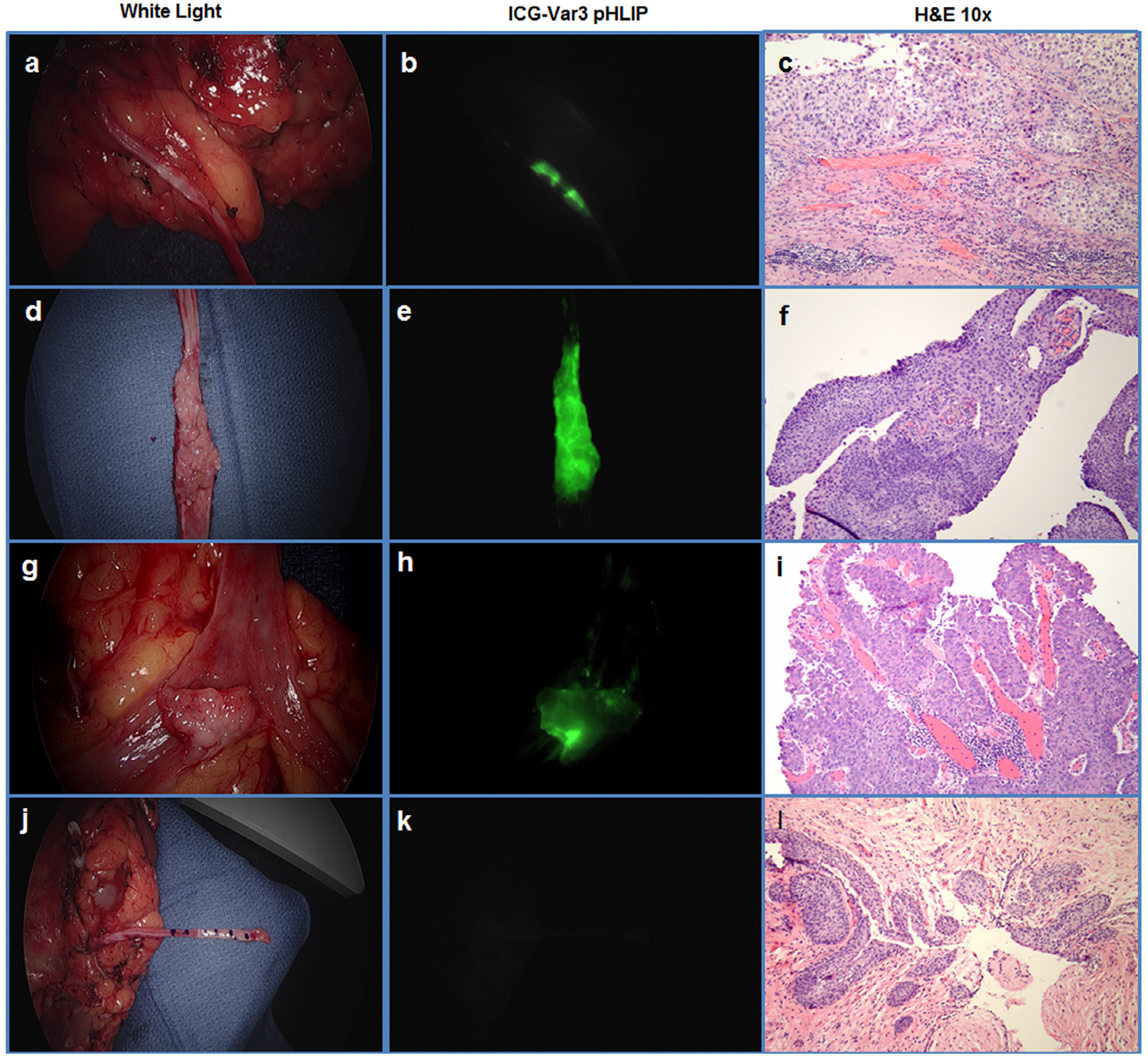
To improve visualization of upper tract urothelial carcinomas. Previous studies using the novel pH low insertion peptide (pHLIP) variant 3 (Var3) conjugated to indocyanine green (ICG) have demonstrated high sensitivity and specificity for imaging of bladder urothelial carcinoma. Here, we describe a novel approach for the imaging of upper tract urothelial carcinomas using ICG-Var3 pHLIP.
Twelve ex-vivo upper urinary tract specimens were irrigated with ICG-Var 3 pHLIP for 15 minutes and then examined using a white light laparoscopic camera followed by near infrared fluorescent (NIRF) imaging using a Stryker 1588 AIM imaging system. Standard histopathologic evaluation was performed and findings were correlated with white light and ICG-Var3 NIRF imaging. One patient who underwent radical nephrectomy for renal cell carcinoma was used as a negative control.
Nineteen lesions were identified on histopathologic evaluation in 10 patients, including 82% high-grade urothelial carcinoma and 18% low-grade urothelial carcinoma. Nineteen (100%) malignant lesions were identified using NIRF imaging, while 15 (78.9%) lesions were identified using conventional white light examination. The sensitivity of ICG-Var3 pHLIP NIRF imaging was 100% compared to 78.9% white light examination. Both modalities are 100% specific. Benign collecting systems and ureters did not show uptake of the pHLIP construct.
In this feasibility study, the ICG-Var3 pHLIP imaging agent demonstrated superior diagnostic performance compared to conventional white light examination. While additional studies are required for validation and in-vivo translation, pHLIP-based imaging represents a promising tool to improve the evaluation and management of upper tract urothelial carcinoma.
提高上尿路尿路上皮癌的可视化程度。先前使用新型 pH 低插入肽 (pHLIP) 变体 3 (Var3) 与吲哚菁绿 (ICG) 缀合的研究已证明对膀胱尿路上皮癌的成像具有高灵敏度和特异性。在这里,我们描述了一种使用 ICG-Var3 pHLIP 对上尿路尿路上皮癌进行成像的新方法。
12 个离体上尿路标本用 ICG-Var3 pHLIP 冲洗 15 分钟,然后用白光腹腔镜相机检查,然后使用 Stryker 1588 AIM 成像系统进行近红外荧光 (NIRF) 成像。进行标准组织病理学评估,并将结果与白光和 ICG-Var3 NIRF 成像相关联。一名接受根治性肾切除术治疗肾细胞癌的患者被用作阴性对照。
在 10 名患者的组织病理学评估中发现了 19 个病变,包括 82%的高级别尿路上皮癌和 18%的低级别尿路上皮癌。19 个(100%)恶性病变通过 NIRF 成像识别,而 15 个(78.9%)病变通过常规白光检查识别。与 78.9%的白光检查相比,ICG-Var3 pHLIP NIRF 成像的灵敏度为 100%。两种方式的特异性均为 100%。良性收集系统和输尿管未摄取 pHLIP 构建体。
在这项可行性研究中,与常规白光检查相比,ICG-Var3 pHLIP 成像剂显示出卓越的诊断性能。虽然还需要进一步的验证和体内转化研究,但基于 pHLIP 的成像代表了一种有前途的工具,可以改善上尿路尿路上皮癌的评估和管理。