Sawant Yogini Nilkantha, Sanghvi Darshana
Department of Radiology, Kokilaben Dhirubhai Ambani Hospital, Mumbai, Maharashtra, India.
Indian J Radiol Imaging. 2018 Oct-Dec;28(4):419-426. doi: 10.4103/ijri.IJRI_77_16.
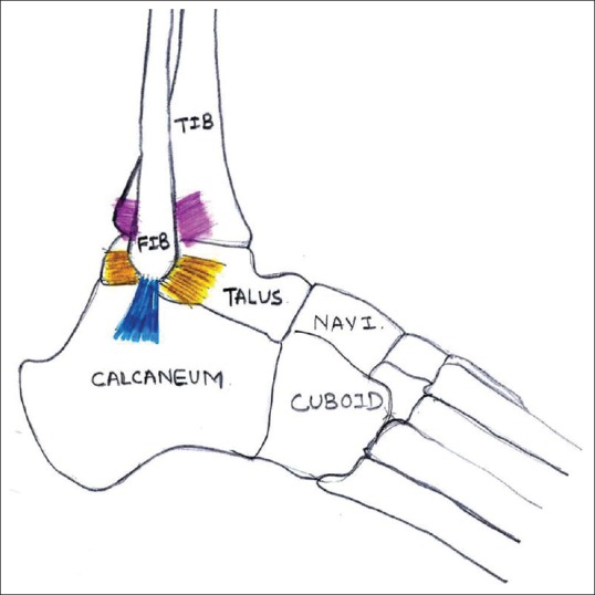
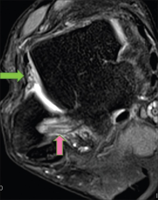
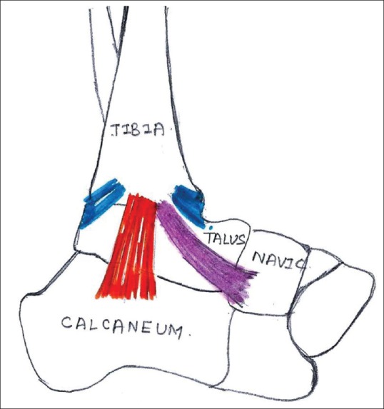
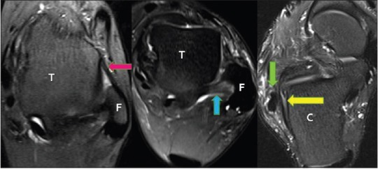
Ankle trauma is commonly encountered and is most often a sprain injury affecting the ligaments. Accurate diagnosis and appropriate treatment rest on knowledge of complex ligamentous anatomy of ankle and the entire spectrum of pathologies. Magnetic resonance imaging (MRI) is the imaging modality of choice for diagnosing ligament pathologies because of its multiplanar capability and high soft tissue contrast. With MRI, it is possible to triage and attribute the cause of post traumatic ankle pain to bone, ligament, or tendon pathologies, which otherwise overlap clinically. In this pictorial essay, emphasis is given to the intricate and unique anatomy and orientation of ankle ligaments. Pathologies of ankle ligaments have been elaborated.
踝关节创伤很常见,最常发生的是影响韧带的扭伤。准确的诊断和恰当的治疗取决于对踝关节复杂韧带解剖结构以及所有病理情况的了解。磁共振成像(MRI)因其多平面成像能力和高软组织对比度,是诊断韧带病变的首选成像方式。借助MRI,可以对创伤后踝关节疼痛的病因进行分类,并将其归因于骨骼、韧带或肌腱病变,否则这些病变在临床上会相互重叠。在这篇图文并茂的文章中,重点介绍了踝关节韧带复杂而独特的解剖结构和方向。文中还阐述了踝关节韧带的病变情况。