Department of Biomedical Engineering, Institute of Basic Medical Sciences and School of Basic Medicine, Peking Union Medical College and Chinese Academy of Medical Sciences, Beijing, China.
Department of Anatomy and Histology, Institute of Basic Medical Sciences and School of Basic Medicine, Peking Union Medical College and Chinese Academy of Medical Sciences, Beijing, China.
Sci Rep. 2019 Jan 22;9(1):253. doi: 10.1038/s41598-018-36319-x.
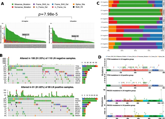
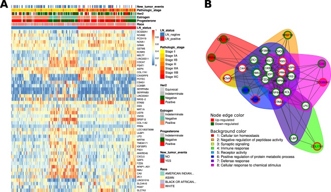
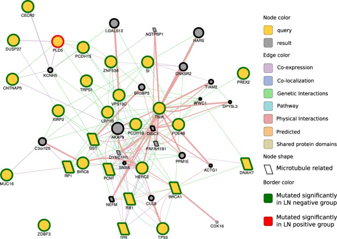
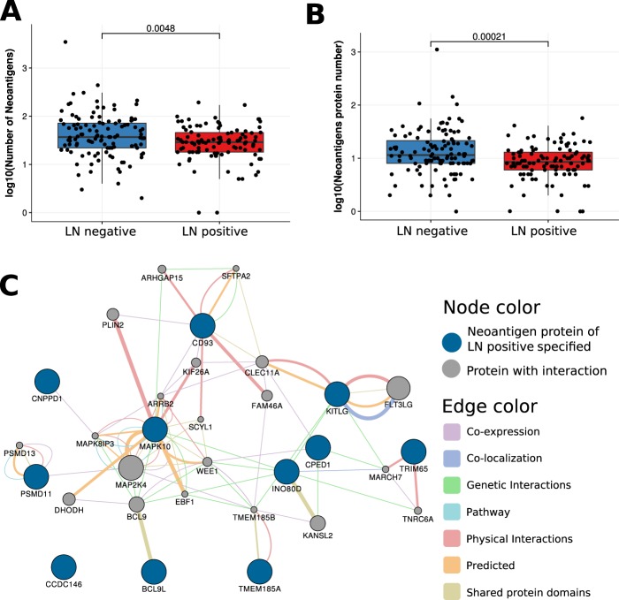
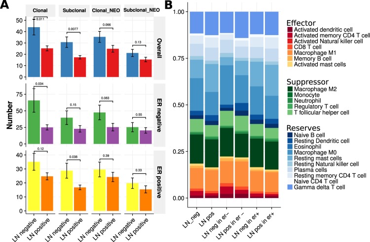
Lymph node metastasis is of major prognostic significance for breast cancer. Lymph node metastasis arises at a very early stage in some patients. Using the data downloaded from the TCGA database, we studied the differences between primary tumors with and without lymph node metastasis at the multi-omics level using bioinformatics approaches. Our study found that low mutation and neoantigen burdens correlated with lymph node metastazation of breast cancer. All three conserved domains in TP53 were mutated in lymph node-negative breast cancers, whereas only one domain was mutated in lymph node-positive samples. Mutations in microtubule-related proteins appear to help immune cells recognize tumors and inhibit their lymph node metastasis. Destroying microtubule-related proteins is a potential therapeutic strategy to inhibit lymph node metastasis of breast cancer. As the neoantigens specifically present in lymph node-positive breast cancers, MAPK10, BC9L, TRIM65, CD93, KITLG, CNPPD1, CPED1, CCDC146, TMEM185A, INO80D, and PSMD11 are potential targets for vaccine design. In the tumor microenvironment, reduced numbers of effector immune cells, especially activated memory CD4+ T cells and activated mast cells, facilitate breast cancer metastasis to the lymph nodes. According to transcriptome data, lymph node metastasis was mostly driven by gene mutation rather than by gene expression. Although differential gene expression analysis was based on lymph node metastasis status, many genes were shown to be differentially expressed based on estrogen receptor status.
淋巴结转移对乳腺癌的预后具有重要意义。在某些患者中,淋巴结转移很早就发生了。我们使用从 TCGA 数据库下载的数据,通过生物信息学方法在多组学水平上研究了有和无淋巴结转移的原发性肿瘤之间的差异。我们的研究发现,低突变和新抗原负担与乳腺癌的淋巴结转移有关。在淋巴结阴性的乳腺癌中,TP53 的三个保守结构域都发生了突变,而在淋巴结阳性的样本中只有一个结构域发生了突变。微管相关蛋白的突变似乎有助于免疫细胞识别肿瘤并抑制其淋巴结转移。破坏微管相关蛋白可能是抑制乳腺癌淋巴结转移的一种潜在治疗策略。MAPK10、BC9L、TRIM65、CD93、KITLG、CNPPD1、CPED1、CCDC146、TMEM185A、INO80D 和 PSMD11 作为仅在淋巴结阳性乳腺癌中特异性存在的新抗原,是疫苗设计的潜在靶点。在肿瘤微环境中,效应免疫细胞数量减少,尤其是激活的记忆 CD4+T 细胞和激活的肥大细胞,促进了乳腺癌向淋巴结的转移。根据转录组数据,淋巴结转移主要由基因突变驱动,而不是基因表达。尽管差异基因表达分析是基于淋巴结转移状态进行的,但许多基因根据雌激素受体状态显示出差异表达。