Tao Pengfei, Feng Jing, Li Qiong, Liu Wei, Yang Lin, Zhao Xiaolong, Ni Huan, Xia Ping
Department of Spine Surgery, Wuhan No. 1 Hospital, Wuhan, Hubei 430022, P.R. China.
Department of Radiology, Taihe Hospital Hubei University of Medicine, Shiyan, Hubei 442000, P.R. China.
Oncol Lett. 2019 Feb;17(2):1791-1797. doi: 10.3892/ol.2018.9739. Epub 2018 Nov 20.
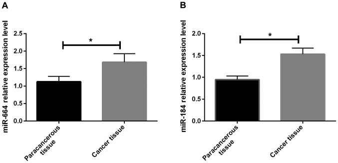
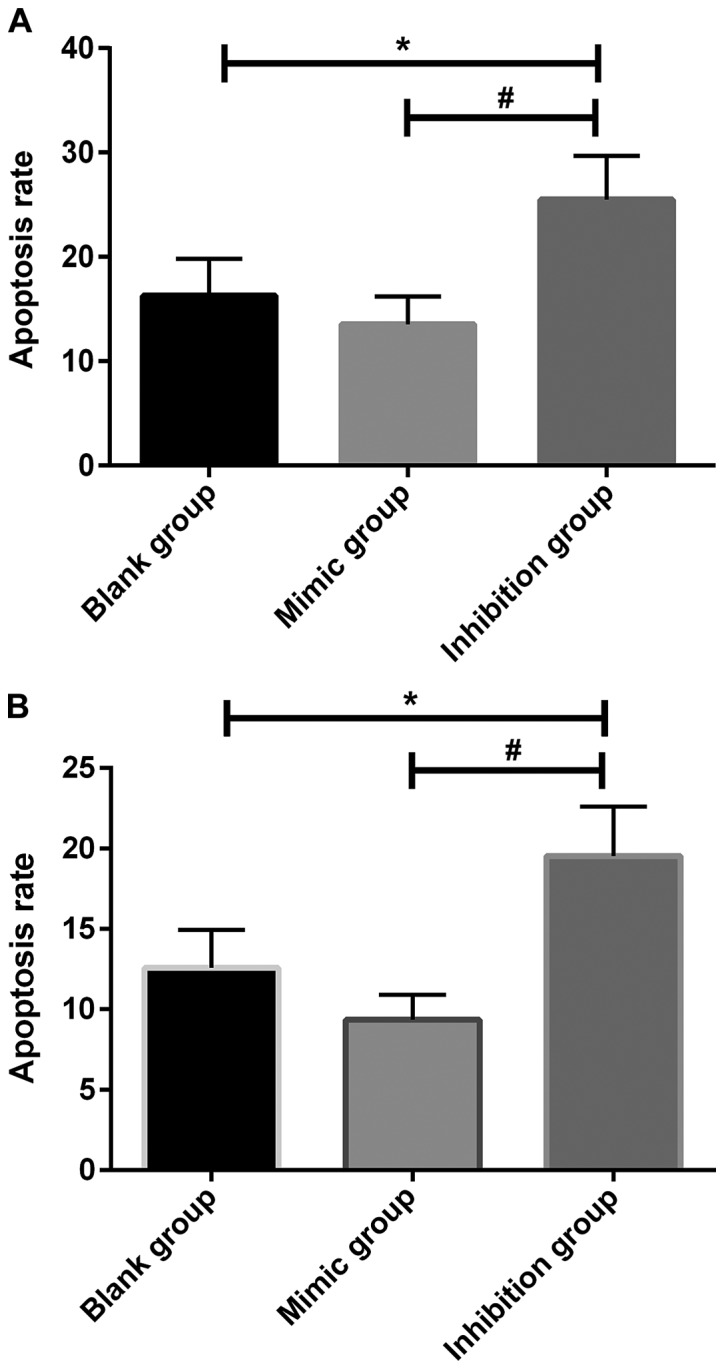
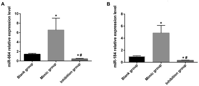
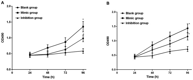
The expression of micro-ribonucleic acid miR-664 and miR-184 on the biological characteristics of osteosarcoma (OS) SOSP-9607 cells was investigated. Eighteen surgical specimens of OS and 18 normal tissue specimens were collected. The expression of miR-664 and miR-184 was detected via fluorescence reverse transcription-quantitative polymerase chain reaction (RT-qPCR). The OS cell line SOSP-9607 was selected as the object of study, and miR-664 blank group, miR-664 mimic group, miR-664 inhibitor group, miR-184 blank group, miR-184 mimic group and miR-184 inhibitor group were established through transfection. Changes in apoptosis were detected via flow cytometry, the cell proliferation capacity was detected via Cell Counting Kit-8 assay, and the cell migration was observed via wound healing assay. In cancer tissues of OS patients, the relative expression of miR-664 and miR-184 was significantly higher than that in para-carcinoma tissues (P<0.05). The cell growth in miR-664 inhibitor group was obviously decreased compared with those in miR-664 blank and mimic groups (P<0.05). There were differences in the cell migration capacity among groups (P<0.01), and the cell scratch areas in miR-664 and miR-184 mimic groups were significantly decreased compared with those in miR-664 and miR-184 blank groups (P<0.05), while they were significantly increased in miR-664 and miR-184 inhibitor groups compared with those in miR-664 and miR-184 blank and mimic groups (P<0.05, P<0.01). There were differences in the apoptosis rate among groups (P<0.01) and apoptosis in miR-664 and miR-184 inhibitor groups was remarkably increased compared with those in miR-664 and miR-184 blank and mimic groups (P<0.05). Downregulating the expression of miR-664 and miR-184 may promote apoptosis, inhibit the proliferation and reduce the migration capacity of SOSP-9607 cells. Therefore, miR-664 and miR-184 may provide a theoretical basis for the target selection in clinical targeted therapy and drug development for OS.
研究了微小核糖核酸miR - 664和miR - 184对骨肉瘤(OS)SOSP - 9607细胞生物学特性的影响。收集18例骨肉瘤手术标本和18例正常组织标本。通过荧光逆转录定量聚合酶链反应(RT - qPCR)检测miR - 664和miR - 184的表达。选取OS细胞系SOSP - 9607作为研究对象,通过转染建立miR - 664空白组、miR - 664模拟物组、miR - 664抑制剂组、miR - 184空白组、miR - 184模拟物组和miR - 184抑制剂组。通过流式细胞术检测细胞凋亡变化,通过细胞计数试剂盒 - 8法检测细胞增殖能力,通过伤口愈合试验观察细胞迁移情况。在OS患者的癌组织中,miR - 664和miR - 184的相对表达明显高于癌旁组织(P<0.05)。与miR - 664空白组和模拟物组相比,miR - 664抑制剂组细胞生长明显降低(P<0.05)。各组细胞迁移能力存在差异(P<0.01),与miR - 664和miR - 184空白组相比,miR - 664和miR - 184模拟物组的细胞划痕面积明显减小(P<0.05),而与miR - 664和miR - 184空白组及模拟物组相比,miR - 664和miR - 184抑制剂组的细胞划痕面积明显增大(P<0.05,P<0.01))。各组凋亡率存在差异(P<0.01),与miR - 664和miR - 184空白组及模拟物组相比,miR - 664和miR - 184抑制剂组的凋亡明显增加(P<0.05)。下调miR - 664和miR - 184的表达可能促进SOSP - 9607细胞凋亡,抑制其增殖并降低其迁移能力。因此,miR - 664和miR - 184可能为骨肉瘤临床靶向治疗和药物研发中的靶点选择提供理论依据。