Department of Rehabilitation Medicine, St. Vincent's Hospital, College of Medicine, The Catholic University of Korea, Seoul, Republic of Korea.
Department of Physical Therapy, College of Public Health & Welfare, The Yongin University, Gyeonggi-do, Republic of Korea.
PLoS One. 2019 Jan 24;14(1):e0210038. doi: 10.1371/journal.pone.0210038. eCollection 2019.
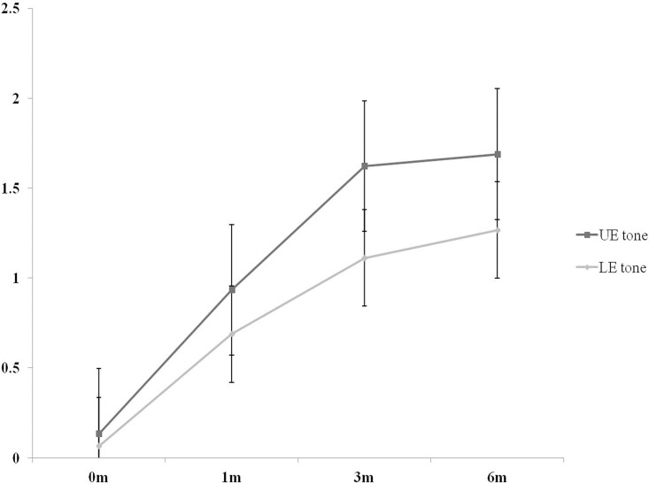
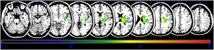
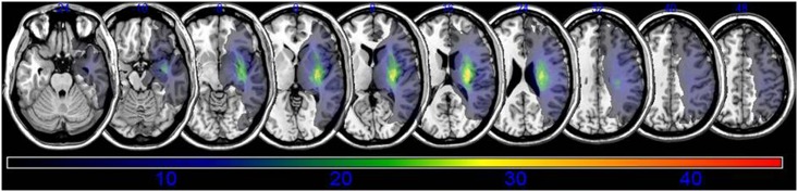
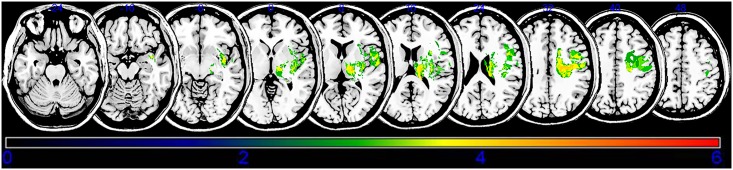
Spasticity is an important barrier that can hinder the restoration of function in stroke patients. Although several studies have attempted to elucidate the relationship between brain lesions and spasticity, the effects of specific brain lesions on the development of spasticity remain unclear. Thus, the present study investigated the effects of stroke lesions on spasticity in stroke patients. The present retrospective longitudinal observational study assessed 45 stroke patients using the modified Ashworth Scale to measure muscle spasticity. Each patient was assessed four times: initially (within 2 weeks of stroke) and at 1, 3, and 6 months after the onset of stroke. Brain lesions were analyzed using voxel-based lesion symptom mapping (VLSM) with magnetic resonance imaging images. Spasticity developed to a certain degree within 3 months in most stroke patients with spasticity. The VLSM method with non-parametric mapping revealed that lesions in the superior corona radiata, posterior limb of the internal capsule, posterior corona radiata, thalamus, putamen, premotor cortex, and insula were associated with the development of upper-limb spasticity. Additionally, lesions of the superior corona radiata, posterior limb of the internal capsule, caudate nucleus, posterior corona radiata, thalamus, putamen, and external capsule were associated with the development of lower-limb spasticity. The present study identified several brain lesions that contributed to post-stroke spasticity. Specifically, the involvement of white matter tracts and the striatum influenced the development of spasticity in the upper and lower limbs of stroke patients. These results may be useful for planning rehabilitation strategies and for understanding the pathophysiology of spasticity in stroke patients.
痉挛是阻碍脑卒中患者功能恢复的重要障碍。尽管已有多项研究试图阐明脑损伤与痉挛之间的关系,但特定脑损伤对痉挛发展的影响仍不清楚。因此,本研究旨在探讨脑卒中患者脑损伤与痉挛的关系。本回顾性纵向观察研究使用改良 Ashworth 量表评估了 45 例脑卒中患者的肌肉痉挛程度。每位患者评估 4 次:脑卒中后 2 周内首次评估,然后分别在 1、3 和 6 个月时进行评估。采用磁共振成像图像的基于体素的病变症状映射(VLSM)分析脑损伤。大多数痉挛性脑卒中患者在 3 个月内痉挛发展到一定程度。非参数映射的 VLSM 方法显示,上放射冠、内囊后肢、后放射冠、丘脑、壳核、运动前皮质和岛叶的病变与上肢痉挛的发展有关。此外,上放射冠、内囊后肢、尾状核、后放射冠、丘脑、壳核和外囊的病变与下肢痉挛的发展有关。本研究确定了几个导致脑卒中后痉挛的脑损伤。具体来说,白质束和纹状体的参与影响了脑卒中患者上下肢痉挛的发展。这些结果可能有助于制定康复策略,并有助于理解脑卒中患者痉挛的病理生理学。