Gandhi Arun, Vellaiyan Subramani, Subramanian Shanmuga, Swamy Shanmugam Thirumalai, Subramanian Kala, Ayyalusamy Anantharaman
Department of Radiation Oncology, Yashoda Hospital, Hyderabad, India.
Research and Development Centre, Bharathiar University, Coimbatore, India. Email:
Asian Pac J Cancer Prev. 2019 Jan 25;20(1):229-234. doi: 10.31557/APJCP.2019.20.1.229.
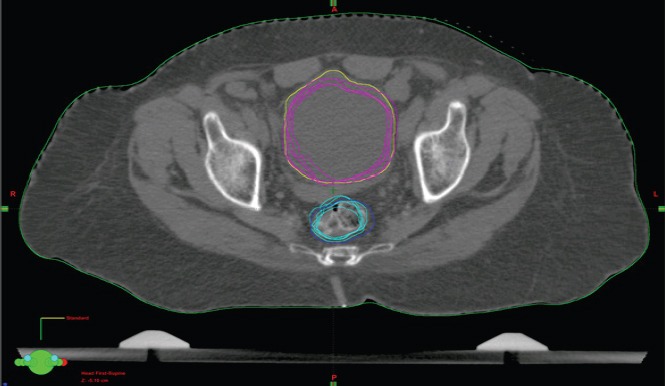
Objective: The purpose of this study is to develop a method to estimate the dose using amorphous silicon detector panel cone beam computed tomography (aSi-kVCBCT) for the OARs and targets in prostate radiotherapy and to compare with the actual planned dose. Methods: The aSi-kVCBCT is used widely in radiotherapy to verify the patient position before treatment. The advancement in aSi-kVCBCT combined with adaptive software allows us to verify the dose distribution in daily acquired CBCT images. CBCT images from 10 patients undergoing radical prostate radiotherapy were included in this study. Patients received total dose of 65Gy in 25 fractions using volumetric modulated arc therapy (VMAT). aSi-kVCBCT scans were acquired before daily treatment and exported to smart adapt software for image adaptation. The planning CT is adapted to daily aSi-kVCBCT images in terms of HU mapping. The primary VMAT plans were copied on to the adapted planning CT images and dose was calculated using Anisotropic Analytic Algorithm (AAA). The DVH is then used to evaluate the volume changes of organs at risk (OAR), the actual dose received by OARs, CTV and PTV during a single fraction. Results: The normalized volume of the bladder and rectum ranged from 0.70–1.66 and 0.70–1.16 respectively. The cumulative mean Sorensen–Dice coefficient values of bladder and rectum were 0.89±0.04 and 0.79±0.06 respectively. The maximum dose differences for CTV and PTV were 2.5% and -4.7% and minimum were 0.1% and 0.1% respectively. Conclusion: The adapted planning CT obtained from daily imaging using aSi-kVCBCT and SmartAdapt® can be used as an effective tool to estimate the volume changes and dose difference in prostate radiotherapy.
本研究的目的是开发一种使用非晶硅探测器平板锥形束计算机断层扫描(aSi-kVCBCT)来估计前列腺放疗中危及器官(OAR)和靶区剂量的方法,并与实际计划剂量进行比较。方法:aSi-kVCBCT在放射治疗中广泛用于治疗前验证患者体位。aSi-kVCBCT与自适应软件的进步使我们能够在每日获取的CBCT图像中验证剂量分布。本研究纳入了10例接受根治性前列腺放疗的患者的CBCT图像。患者使用容积调强弧形放疗(VMAT),25次分割接受总剂量65Gy。在每日治疗前进行aSi-kVCBCT扫描,并导出到智能自适应软件进行图像适配。在HU映射方面,将计划CT适配到每日aSi-kVCBCT图像。将初始VMAT计划复制到适配后的计划CT图像上,并使用各向异性解析算法(AAA)计算剂量。然后使用剂量体积直方图(DVH)评估单分次期间危及器官(OAR)、CTV和PTV的体积变化、OAR实际接受的剂量。结果:膀胱和直肠的归一化体积分别为0.70 - 1.66和0.70 - 1.16。膀胱和直肠的累积平均索伦森 - 戴斯系数值分别为0.89±0.04和0.79±