Schrooten Maarten, Vandenberghe Rik, Peeters Ronald, Dupont Patrick
Laboratory for Cognitive Neurology, KU Leuven, Leuven, Belgium.
Department of Neurology, University Hospitals Leuven, Leuven, Belgium.
Front Neurosci. 2019 Jan 10;12:1009. doi: 10.3389/fnins.2018.01009. eCollection 2018.
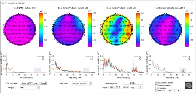
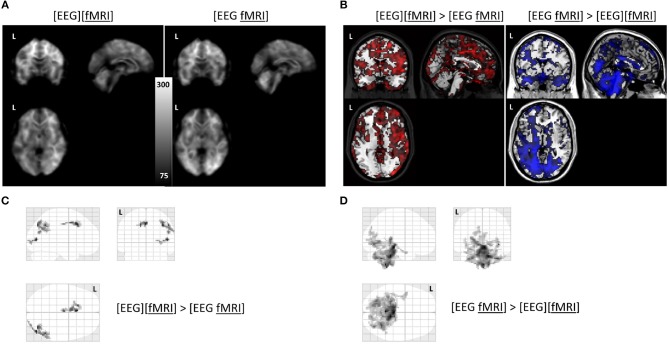
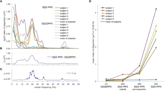
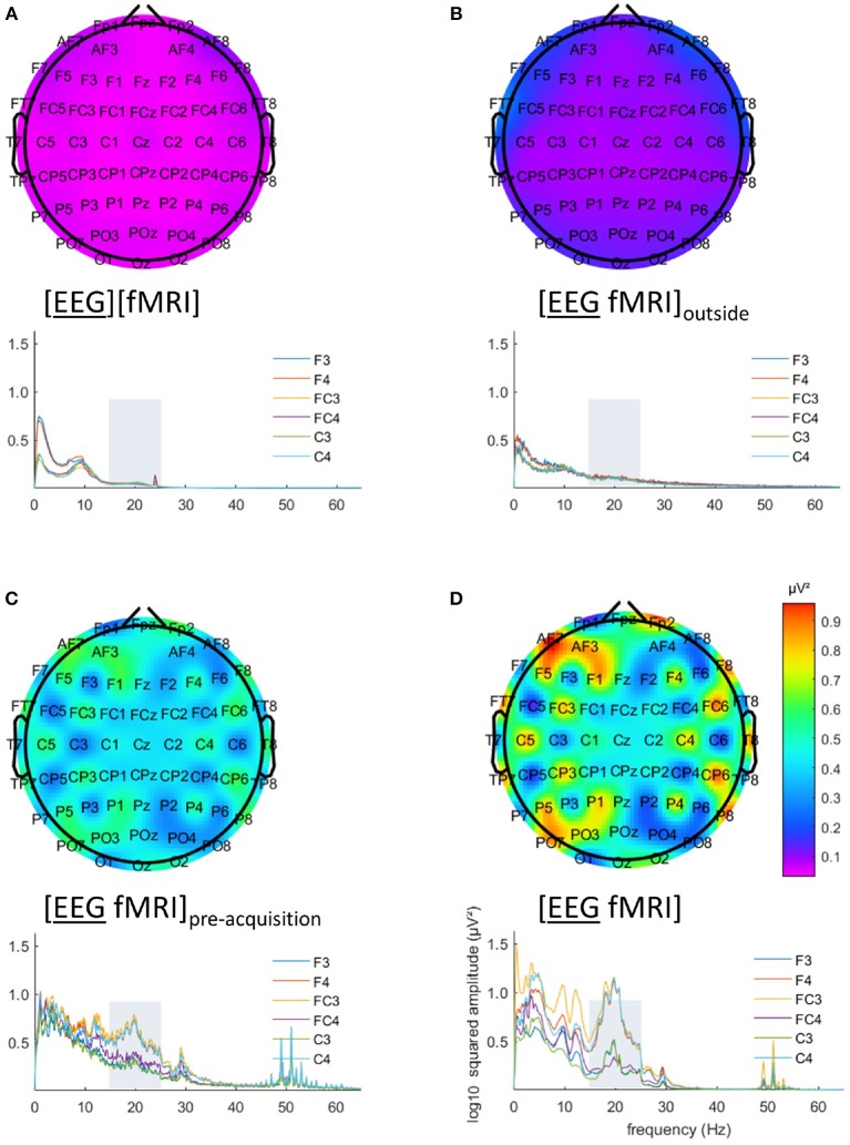
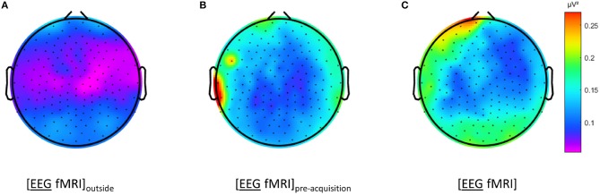
Simultaneous registration of scalp electroencephalography (EEG) and functional magnetic resonance imaging (fMRI) is considered an attractive approach for studying brain function non-invasively. It combines the better spatial resolution of fMRI with the better temporal resolution of EEG, but comes at the cost of increased measurement artifact and the accompanying artifact preprocessing. This paper presents a study of the residual signal quality based on temporal signal to noise ratio (TSNR) for fMRI and fast Fourier transform (FFT) for EEG, after optimized conventional signal preprocessing. Measurements outside the magnetic resonance imaging scanner and inside the scanner prior to and during image acquisition were compared. For EEG, frequency and region dependent significant effects on FFT squared amplitudes were observed between separately vs. simultaneously recorded EEG and fMRI, with larger effects during image acquisition than without image acquisition inside the scanner bore. A graphical user interface was developed to aid in quality checking these measurements. For fMRI, separately recorded EEG-fMRI revealed relatively large areas with a significantly higher TSNR in right occipital and parietal regions and in the cingulum, compared to separately recorded EEG-fMRI. Simultaneously recorded EEG-fMRI showed significantly higher TSNR in inferior occipital cortex, diencephalon and brainstem, compared to separately recorded EEG-fMRI. Quantification of EEG and fMRI signals showed significant, but sometimes subtle, changes between separate compared to simultaneous EEG-fMRI measurements. To avoid interference with the experiment of interest in a simultaneous EEG-fMRI measurement, it seems warranted to perform a quantitative evaluation to ensure that there are no such uncorrectable effects present in regions or frequencies that are of special interest to the research question at hand.
同步记录头皮脑电图(EEG)和功能磁共振成像(fMRI)被认为是一种用于无创研究脑功能的有吸引力的方法。它将fMRI更好的空间分辨率与EEG更好的时间分辨率结合起来,但代价是测量伪影增加以及随之而来的伪影预处理。本文介绍了一项基于fMRI的时间信噪比(TSNR)和EEG的快速傅里叶变换(FFT)对优化后的传统信号预处理后的残余信号质量的研究。比较了在磁共振成像扫描仪外部以及在扫描仪内部图像采集之前和期间的测量结果。对于EEG,在分别记录的EEG与fMRI以及同时记录的EEG与fMRI之间,观察到对FFT平方幅度的频率和区域依赖性显著影响,在扫描仪孔内进行图像采集时的影响比不进行图像采集时更大。开发了一个图形用户界面来辅助检查这些测量的质量。对于fMRI,与单独记录的EEG-fMRI相比,单独记录的EEG-fMRI显示右枕叶和顶叶区域以及扣带中有相对较大的区域具有显著更高的TSNR。与单独记录的EEG-fMRI相比,同时记录的EEG-fMRI显示枕下回、间脑和脑干中的TSNR显著更高。EEG和fMRI信号的量化显示,与同时进行的EEG-fMRI测量相比,单独测量之间存在显著但有时细微的变化。为了避免在同步EEG-fMRI测量中干扰感兴趣的实验,似乎有必要进行定量评估,以确保在手头研究问题特别感兴趣的区域或频率中不存在此类无法纠正的影响。