Salvador João Cunha, Coutinho Mónica Portela, Venâncio José Marques, Viamonte Bárbara
Department of Radiology, Instituto Português de Oncologia de Lisboa Francisco Gentil, Rua Prof. Lima Basto, Lisboa, Portugal.
Department of Radiology, Hospital Universitário de São João, Centro Hospitalar São João, Porto, Portugal.
Insights Imaging. 2019 Jan 28;10(1):4. doi: 10.1186/s13244-019-0687-9.
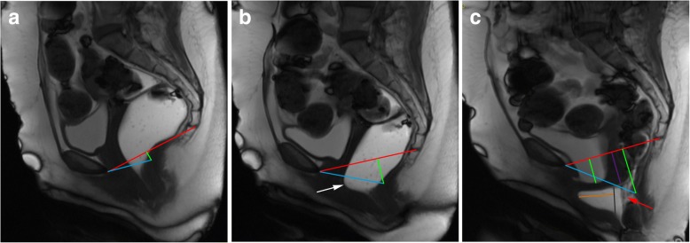
Pelvic floor dysfunctions represent a range of functional disorders that frequently occur in adult women, carrying a significant burden on the quality of life, and its incidence tends to increase attending to the expected aging of the population. Pelvic floor dysfunctions can manifest as incontinence, constipation, and prolapsed pelvic organs. Since pelvic floor weakness is frequently generalized and clinically underdiagnosed, imaging evaluation is of major importance, especially prior to surgical correction. Given some interobserver variability of soft-tissue measurements, MR defecography allows a noninvasive, radiation-free, multiplanar dynamic evaluation of the three pelvic compartments simultaneously and with high spatial and temporal resolution. Both static/anatomic and dynamic/functional findings are important, since pelvic disorders can manifest as whole pelvic floor weakness/dysfunction or as an isolated or single compartment disorder. Imaging has a preponderant role in accessing pelvic floor disorders, and dynamic MR defecography presents as a reliable option, being able to evaluate the entire pelvic floor for optimal patient management before surgery. The purpose of this article is to address the female pelvic anatomy and explain the appropriate MR Defecography protocol, along with all the anatomic points, lines, angles, and measurements needed for a correct interpretation, to later focus on the different disorders of the female pelvic floor, illustrated with MR defecography images, highlighting the role of this technique in accessing these pathologic conditions.
盆底功能障碍是一系列常见于成年女性的功能性疾病,给生活质量带来了重大负担,并且随着人口预期寿命的增加,其发病率有上升趋势。盆底功能障碍可表现为尿失禁、便秘和盆腔器官脱垂。由于盆底肌无力常常是全身性的且在临床上易被漏诊,影像学评估尤为重要,特别是在手术矫正之前。鉴于软组织测量存在一定的观察者间差异,磁共振排粪造影能够对三个盆腔腔室进行无创、无辐射的多平面动态评估,且具有高空间和时间分辨率。静态/解剖学和动态/功能性表现均很重要,因为盆腔疾病可表现为整个盆底肌无力/功能障碍,也可表现为孤立的或单一腔室的疾病。影像学在诊断盆底疾病中起着重要作用,动态磁共振排粪造影是一种可靠的选择,能够在手术前对整个盆底进行评估,以实现对患者的最佳管理。本文旨在阐述女性盆腔解剖结构,解释合适的磁共振排粪造影方案,以及正确解读所需的所有解剖学要点、线条、角度和测量方法,随后聚焦于女性盆底的不同疾病,并配以磁共振排粪造影图像进行说明,突出该技术在诊断这些病理状况中的作用。