Kruppa Michael D, Jacobs Jeremy, King-Hook Kelsey, Galloway Keleigh, Berry Amy, Kintner Jennifer, Whittimore Judy D, Fritz Rolf, Schoborg Robert V, Hall Jennifer V
Department of Biomedical Sciences, Quillen College of Medicine, East Tennessee State University, Johnson City, TN, United States.
Center for Infectious Disease, Inflammation and Immunity, Quillen College of Medicine, East Tennessee State University, Johnson City, TN, United States.
Front Microbiol. 2019 Jan 14;9:3270. doi: 10.3389/fmicb.2018.03270. eCollection 2018.
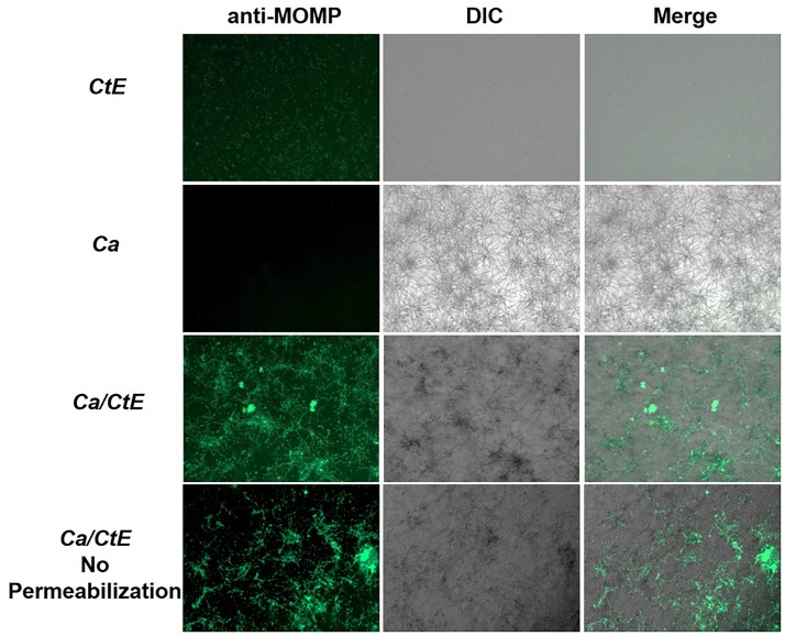
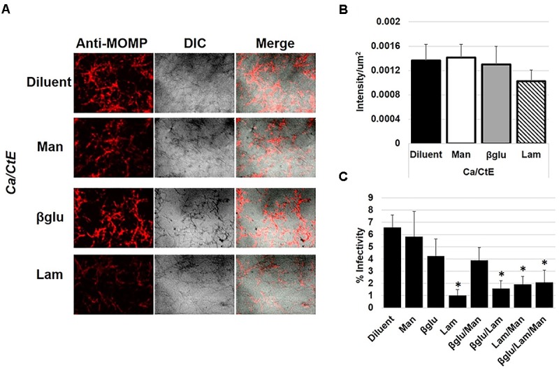
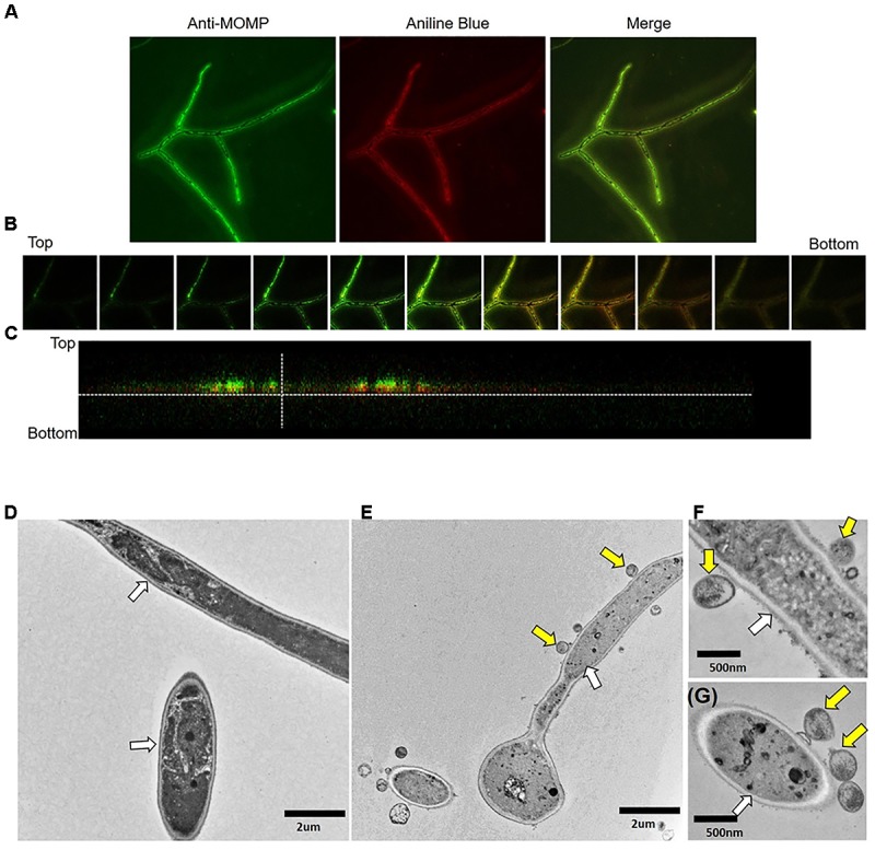
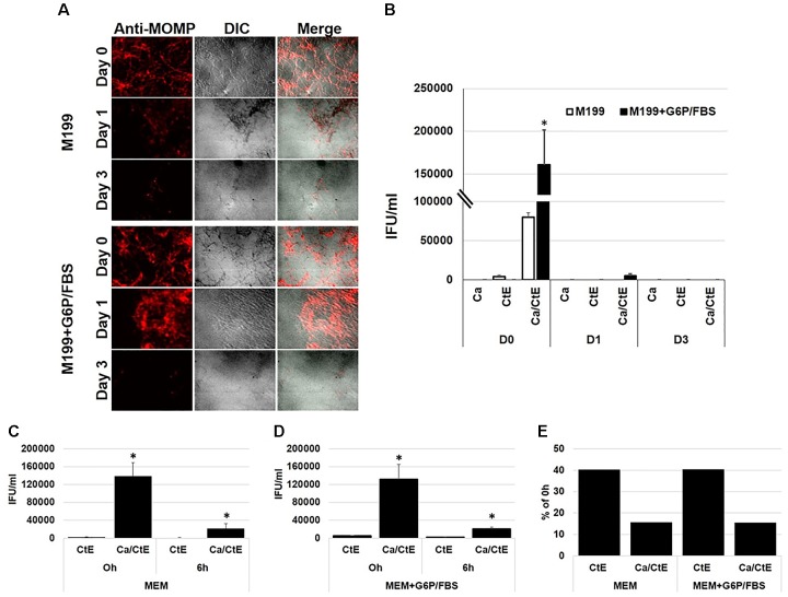
Microbial interactions represent an understudied facet of human health and disease. In this study, the interactions that occur between and the opportunistic fungal pathogen, were investigated. is a common component of the oral and vaginal microbiota responsible for thrush and vaginal yeast infections. Normally, exist in the body as yeast. However, disruptions to the microbiota create conditions that allow expanded growth of , conversion to the hyphal form, and tissue invasion. Previous studies have shown that a myriad of outcomes can occur when interacts with pathogenic bacteria. To determine if physically interacts with , we incubated chlamydial elementary bodies (EB) in medium alone or with yeast or hyphal forms for 1 h. Following incubation, the samples were formaldehyde-fixed and processed for immunofluorescence assays using anti-chlamydial MOMP or anti- chlamydial LPS antibodies. Replicate samples were replenished with culture medium and incubated at 35°C for 0-120 h prior to fixation for immunofluorescence analysis or collection for EB infectivity assays. Data from this study indicates that both serovar E and EB bind to yeast and hyphal forms. This interaction was not blocked by pre-incubation of EB with the cell wall components, mannan or β-glucans, suggesting that EB interact with a cell wall protein or other structure. Bound EB remained attached to for a minimum of 5 days (120 h). Infectivity assays demonstrated that EB bound to are infectious immediately following binding (0h). However, once bound to , EB infectivity decreased at a faster rate than EB in medium alone. At 6h post binding, 40% of EB incubated in medium alone remained infectious compared to only 16% of EB bound to . Likewise, pre-incubation of EB with laminarin, a soluble preparation of β-glucan, alone or in combination with other fungal cell wall components significantly decreases chlamydial infectivity in HeLa cells. These data indicate that interactions between EB and inhibit chlamydial infectivity, possibly by physically blocking EB interactions with host cell receptors.
微生物相互作用是人类健康与疾病中一个研究较少的方面。在本研究中,对[未提及的微生物]与机会性真菌病原体[白色念珠菌]之间发生的相互作用进行了研究。白色念珠菌是口腔和阴道微生物群的常见组成部分,可导致鹅口疮和阴道酵母菌感染。通常情况下,白色念珠菌以酵母形式存在于体内。然而,微生物群的破坏会创造条件,使白色念珠菌得以大量生长、转化为菌丝形式并侵入组织。先前的研究表明,当[未提及的微生物]与病原菌相互作用时,会出现多种结果。为了确定[未提及的微生物]是否与白色念珠菌发生物理相互作用,我们将衣原体原体(EB)单独置于培养基中,或与白色念珠菌的酵母或菌丝形式一起孵育1小时。孵育后,将样品用甲醛固定,并使用抗衣原体主要外膜蛋白(MOMP)或抗衣原体脂多糖(LPS)抗体进行免疫荧光检测。重复样品补充培养基后,在35°C孵育0 - 120小时,然后固定用于免疫荧光分析或收集用于EB感染性测定。本研究的数据表明,血清型E的[未提及的微生物]和衣原体EB都能与白色念珠菌的酵母和菌丝形式结合。这种相互作用不会因将EB与白色念珠菌细胞壁成分甘露聚糖或β - 葡聚糖预孵育而被阻断,这表明EB与白色念珠菌细胞壁蛋白或其他结构相互作用。结合的EB至少在5天(120小时)内仍附着在白色念珠菌上。感染性测定表明,与白色念珠菌结合的EB在结合后立即具有感染性(0小时)。然而,一旦与白色念珠菌结合,EB的感染性下降速度比单独在培养基中的EB更快。结合后6小时,单独在培养基中孵育的EB中有40%仍具有感染性,而与白色念珠菌结合的EB只有16%具有感染性。同样,将EB与β - 葡聚糖的可溶性制剂海带多糖单独或与其他真菌细胞壁成分一起预孵育,会显著降低衣原体在HeLa细胞中的感染性。这些数据表明,EB与白色念珠菌之间的相互作用可能通过物理阻断EB与宿主细胞受体的相互作用来抑制衣原体的感染性。