Department of Human Neurosciences, Sapienza University of Rome, Rome, Italy.
Multiple Sclerosis Centre, Sant'Andrea Hospital, Sapienza University of Rome, Rome, Italy.
Neural Plast. 2018 Dec 31;2018:3419871. doi: 10.1155/2018/3419871. eCollection 2018.
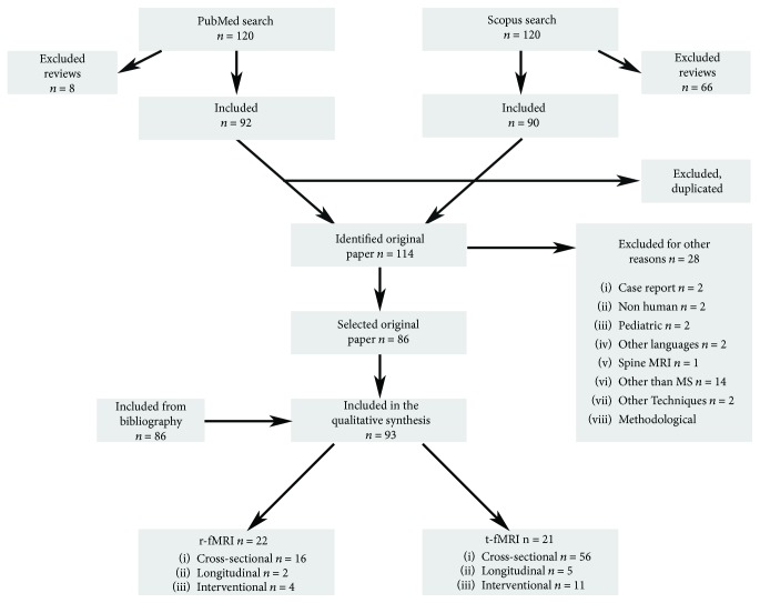
Neuroplasticity, which is the ability of the brain to adapt to internal and external environmental changes, physiologically occurs during growth and in response to damage. The brain's response to damage is of particular interest in multiple sclerosis, a chronic disease characterized by inflammatory and neurodegenerative damage to the central nervous system. Functional MRI (fMRI) is a tool that allows functional changes related to the disease and to its evolution to be studied in vivo. Several studies have shown that abnormal brain recruitment during the execution of a task starts in the early phases of multiple sclerosis. The increased functional activation during a specific task observed has been interpreted mainly as a mechanism of adaptive plasticity designed to contrast the increase in tissue damage. More recent fMRI studies, which have focused on the activity of brain regions at rest, have yielded nonunivocal results, suggesting that changes in functional brain connections represent mechanisms of either adaptive or maladaptive plasticity. The few longitudinal studies available to date on disease evolution have also yielded discrepant results that are likely to depend on the clinical features considered and the length of the follow-up. Lastly, fMRI has been used in interventional studies to investigate plastic changes induced by pharmacological therapy or rehabilitation, though whether such changes represent a surrogate of neuroplasticity remains unclear. The aim of this paper is to systematically review the existing literature in order to provide an overall description of both the neuroplastic process itself and the evolution in the use of fMRI techniques as a means of assessing neuroplasticity. The quantitative and qualitative approach adopted here ensures an objective analysis of published, peer-reviewed research and yields an overview of up-to-date knowledge.
神经可塑性是大脑适应内部和外部环境变化的能力,在生长过程中和对损伤的反应中生理上发生。大脑对损伤的反应在多发性硬化症中特别有趣,多发性硬化症是一种以中枢神经系统炎症和神经退行性损伤为特征的慢性疾病。功能磁共振成像 (fMRI) 是一种工具,可用于研究与疾病及其演变相关的功能变化。多项研究表明,在执行任务期间,大脑的异常募集始于多发性硬化症的早期阶段。在特定任务期间观察到的功能激活增加主要被解释为一种适应可塑性机制,旨在抵消组织损伤的增加。最近的 fMRI 研究集中在静息状态下的脑区活动,得出了不一致的结果,这表明功能脑连接的变化代表了适应或适应不良可塑性的机制。迄今为止,关于疾病演变的少数纵向研究也得出了不一致的结果,这些结果可能取决于所考虑的临床特征和随访时间。最后,fMRI 已用于干预性研究,以研究药物治疗或康复引起的可塑性变化,尽管这些变化是否代表神经可塑性的替代物尚不清楚。本文的目的是系统地回顾现有文献,以便全面描述神经可塑性过程本身以及 fMRI 技术在评估神经可塑性方面的演变。采用的定量和定性方法确保了对已发表的同行评议研究的客观分析,并概述了最新知识。