Nie Xin, Liu Cong, Guo Qian, Zheng Ming-Jun, Gao Ling-Ling, Li Xiao, Liu Da-Wo, Zhu Lian-Cheng, Liu Juan-Juan, Lin Bei
Department of Obstetrics and Gynaecology, Shengjing Hospital Affiliated to China Medical University, Liaoning, China,
Key Laboratory of Maternal-Fetal Medicine of Liaoning Province, Key Laboratory of Obstetrics and Gynecology of Higher Education of Liaoning Province, Liaoning, China,
Cancer Manag Res. 2019 Jan 17;11:839-855. doi: 10.2147/CMAR.S186080. eCollection 2019.
Transmembrane protein with epidermal growth factor-like and two follistatin-like domains 1 (TMEFF1) has an anticarcinogenic effect in brain tumors. However, little is known about the role of TMEFF1 in epithelial ovarian cancer (EOC).
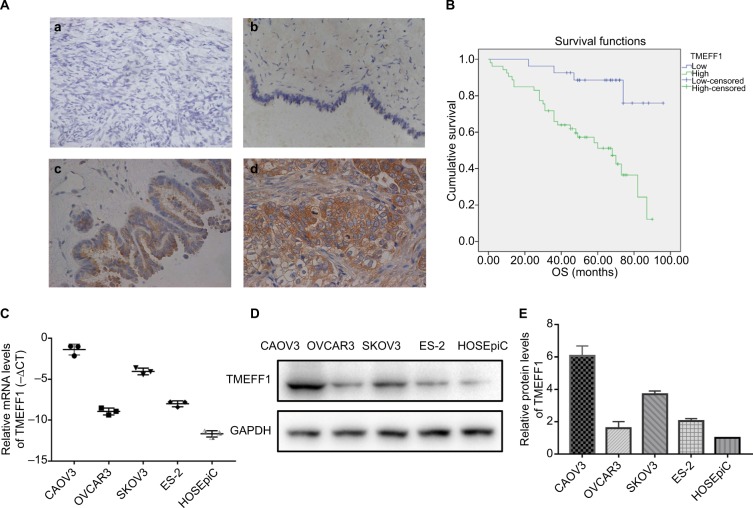
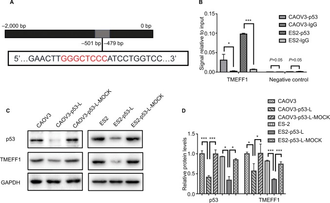
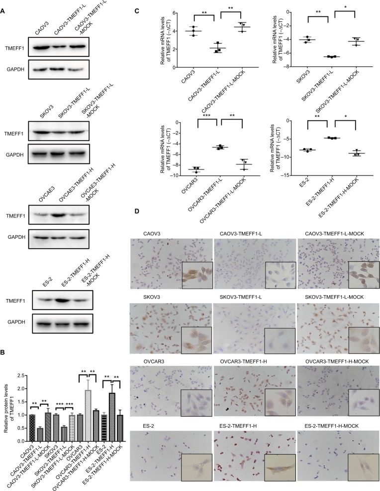
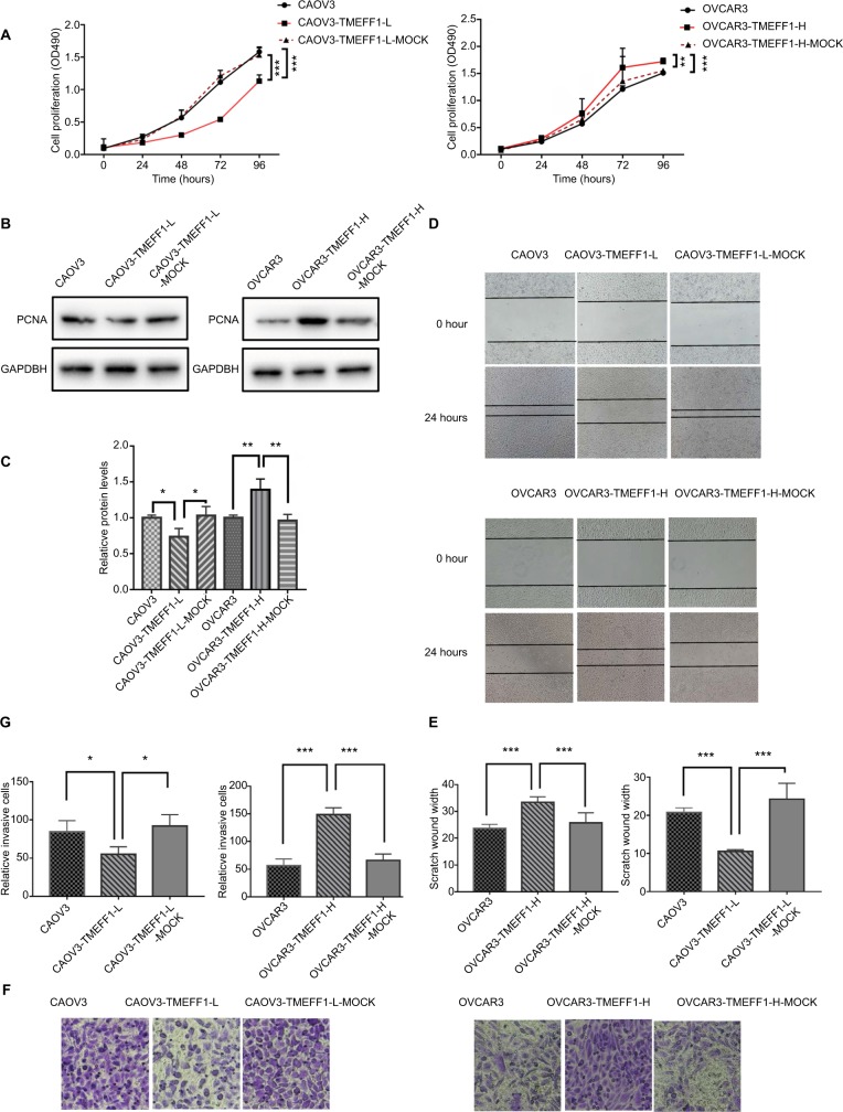
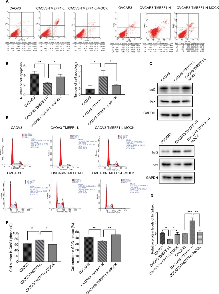
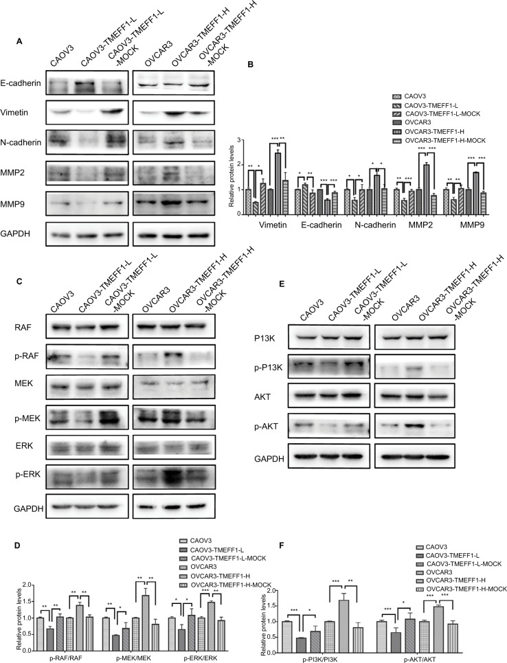
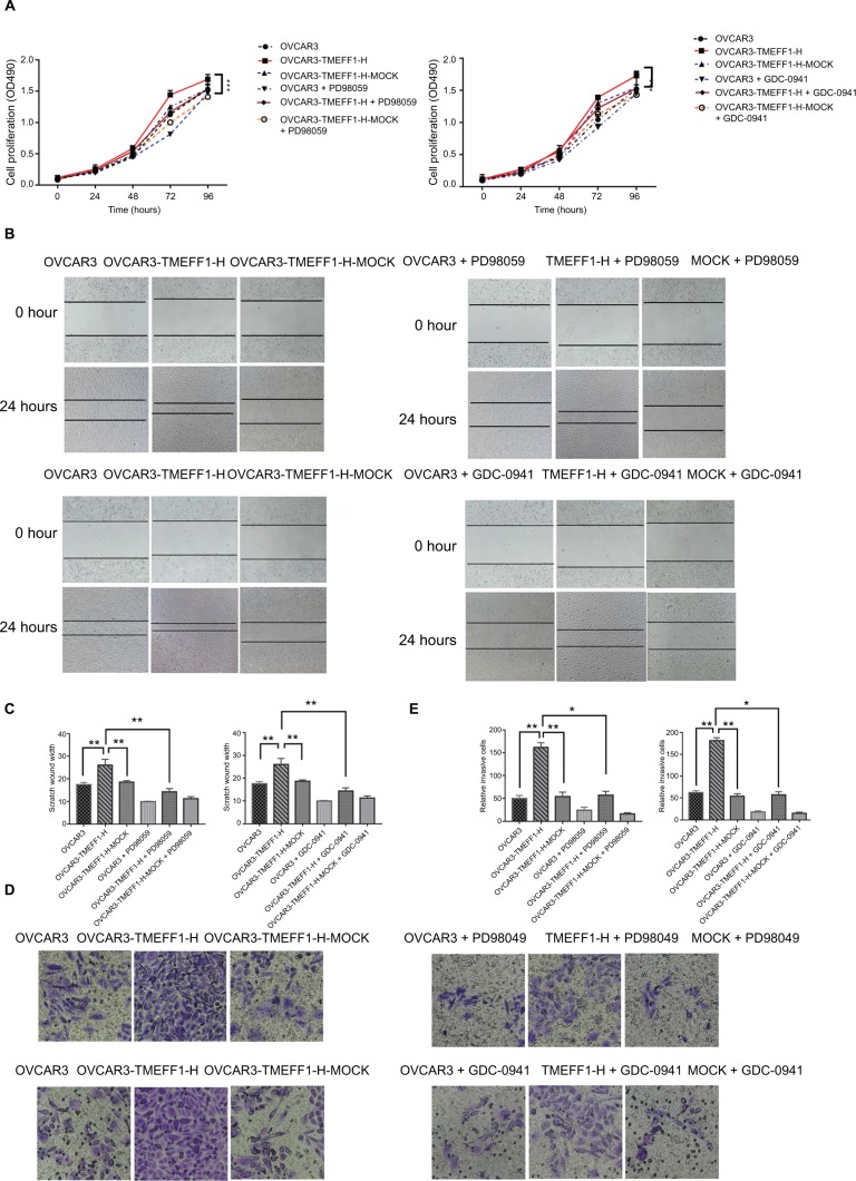
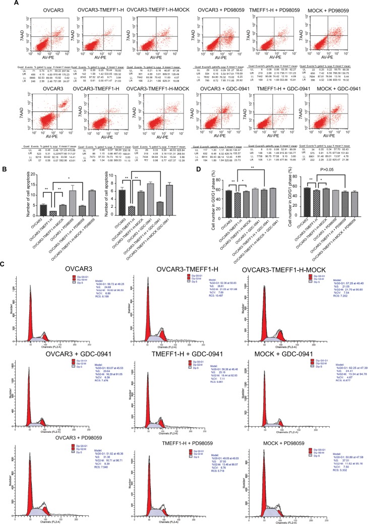
TMEFF1 expression in EOC was detected by immunohistochemistry; its relationship with clinical pathological parameters and its influence on prognosis were analyzed. The MTT, scratch, Transwell assays, and flow cytometry were used to assess the malignant behavior of ovarian cancer cell. Changes in node proteins in MAPK and PI3K/AKT signaling pathways and the expression of epithelial-mesenchymal transformation markers were measured by Western blot. The regulatory effect of p53 on TMEFF1 was verified by chromatin immunoprecipitation (ChIP) assay and Western blot.
TMEFF1 expression was higher in the EOC group than in the borderline and benign tumor groups and normal ovary group; its high expression was significantly related to International Federation of Gynecology and Obstetrics stage (=0.024) and independently predicted shorter overall survival (<0.01). TMEFF1 overexpression in ovarian cancer cells induced increased cellular proliferation, migration, and invasion but reduced apoptosis. In addition, the percentage of phosphorylated node proteins in MAPK and PI3K/AKT signaling pathways increased significantly. The expression of E-cadherin decreased but that of vimentin and N-cadherin increased. After the addition of MAPK (PD98059) and PI3K (GDC-0941) pathway inhibitors, ovarian cancer cells overexpressing TMEFF1 showed suppressed malignant behavior. TMEFF1 protein expression in an ovarian cancer cell lines (CAOV3 and ES-2) was downregulated after the inhibition of . The transcription factor, p53, bound the promoter region of the gene according to ChIP.
is a carcinogenic gene in ovarian cancer and can be regulated by p53 transcription. Through MAPK and PI3K/AKT signaling pathways, TMEFF1 promotes the malignant behavior in EOC. Therefore, TMEFF1 may be considered as a potential therapeutic target for ovarian cancer.
具有表皮生长因子样和两个卵泡抑素样结构域的跨膜蛋白1(TMEFF1)在脑肿瘤中具有抗癌作用。然而,关于TMEFF1在上皮性卵巢癌(EOC)中的作用知之甚少。
采用免疫组织化学法检测EOC中TMEFF1的表达;分析其与临床病理参数的关系及其对预后的影响。采用MTT法、划痕实验、Transwell实验和流式细胞术评估卵巢癌细胞的恶性行为。通过蛋白质免疫印迹法检测MAPK和PI3K/AKT信号通路中节点蛋白的变化以及上皮-间质转化标志物的表达。通过染色质免疫沉淀(ChIP)实验和蛋白质免疫印迹法验证p53对TMEFF1的调控作用。
EOC组中TMEFF1的表达高于交界性肿瘤组、良性肿瘤组和正常卵巢组;其高表达与国际妇产科联盟分期显著相关(=0.024),并独立预测总生存期较短(<0.01)。卵巢癌细胞中TMEFF1的过表达导致细胞增殖、迁移和侵袭增加,但凋亡减少。此外,MAPK和PI3K/AKT信号通路中磷酸化节点蛋白的百分比显著增加。E-钙黏蛋白的表达降低,而波形蛋白和N-钙黏蛋白的表达增加。添加MAPK(PD98059)和PI3K(GDC-0941)信号通路抑制剂后,过表达TMEFF1的卵巢癌细胞的恶性行为受到抑制。在卵巢癌细胞系(CAOV3和ES-2)中,抑制 后TMEFF1蛋白表达下调。根据ChIP实验,转录因子p53与 基因的启动子区域结合。
是卵巢癌中的致癌基因,可受p53转录调控。通过MAPK和PI3K/AKT信号通路,TMEFF1促进EOC中的恶性行为。因此,TMEFF1可被视为卵巢癌的潜在治疗靶点。