• 文献检索
  • 文档翻译
  • 深度研究
  • 学术资讯
  • Suppr Zotero 插件Zotero 插件
  • 邀请有礼
  • 套餐&价格
  • 历史记录
应用&插件
Suppr Zotero 插件Zotero 插件浏览器插件Mac 客户端Windows 客户端微信小程序
定价
高级版会员购买积分包购买API积分包
服务
文献检索文档翻译深度研究API 文档MCP 服务
关于我们
关于 Suppr公司介绍联系我们用户协议隐私条款
关注我们

Suppr 超能文献

核心技术专利:CN118964589B侵权必究
粤ICP备2023148730 号-1Suppr @ 2026

文献检索

告别复杂PubMed语法,用中文像聊天一样搜索,搜遍4000万医学文献。AI智能推荐,让科研检索更轻松。

立即免费搜索

文件翻译

保留排版,准确专业,支持PDF/Word/PPT等文件格式,支持 12+语言互译。

免费翻译文档

深度研究

AI帮你快速写综述,25分钟生成高质量综述,智能提取关键信息,辅助科研写作。

立即免费体验

临床实践中胸腺影像学与重症肌无力的相关性

Correlation Between Thymus Radiology and Myasthenia Gravis in Clinical Practice.

作者信息

Luo Huan, Xie Shanshan, Ma Chao, Zhang Wenqiang, Tschöpe Carsten, Fa Xianen, Cheng Jingliang, Cao Jing

机构信息

MR Department, The First Affiliated Hospital of Zhengzhou University, Zhengzhou, China.

Department of Ophthalmology, Campus Virchow, Charité - Universitätsmedizin Berlin, Berlin, Germany.

出版信息

Front Neurol. 2019 Jan 15;9:1173. doi: 10.3389/fneur.2018.01173. eCollection 2018.

DOI:10.3389/fneur.2018.01173
PMID:30697185
原文链接:https://pmc.ncbi.nlm.nih.gov/articles/PMC6340958/
Abstract

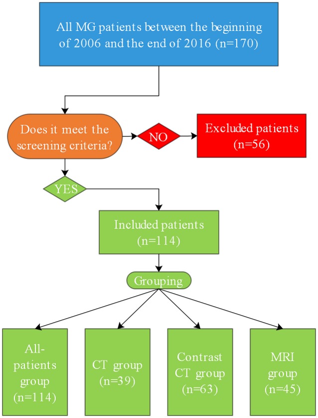
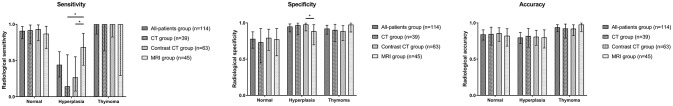
The ability to distinguish between a normal thymus, thymic hyperplasia, and thymoma should aid in clinical management and decision making for patients with myasthenia gravis (MG). We sought to determine the accuracy of routine radiological examinations in predicting thymic pathology. We retrospectively analyzed the records of patients with MG who had undergone thymectomy from the Second Affiliated Hospital of Zhengzhou University. Each patient received at least one initial radiological diagnosis and one histological diagnosis, and the patients were classified into the all-patient, CT, contrast CT, and MRI groups. The sensitivity, accuracy and specificity of each group were calculated for different histological types. This study included 114 patients. All sensitivity, specificity and accuracy values except for sensitivity to hyperplasia in each group for different histological types were satisfactory. MRI had higher sensitivity (68.4, 95% CI: 43.5-87.4%) to histological hyperplasia than did CT (14.3, 95% CI: 0.4-57.9%) and contrast CT (26.7, 95% CI: 7.8-55.1%). Contrast CT had higher specificity (97.9, 95% CI: 88.9-99.95%) for histological hyperplasia than did MRI (88.5, 95% CI: 69.9-97.6%). For patients with MG, CT, contrast CT, and MRI examinations can effectively identify thymoma. Additionally, compared with CT or contrast CT, MRI may have a stronger ability to distinguish thymoma and detect hyperplasia.

摘要

区分正常胸腺、胸腺增生和胸腺瘤的能力有助于重症肌无力(MG)患者的临床管理和决策。我们试图确定常规放射学检查在预测胸腺病理方面的准确性。我们回顾性分析了郑州大学第二附属医院接受胸腺切除术的MG患者的记录。每位患者至少接受一次初始放射学诊断和一次组织学诊断,并将患者分为全患者组、CT组、增强CT组和MRI组。计算每组针对不同组织学类型的敏感性、准确性和特异性。本研究纳入了114例患者。除了每组针对不同组织学类型对增生的敏感性外,所有敏感性、特异性和准确性值均令人满意。MRI对组织学增生的敏感性(68.4,95%CI:43.5-87.4%)高于CT(14.3,95%CI:0.4-57.9%)和增强CT(26.7,95%CI:7.8-55.1%)。增强CT对组织学增生的特异性(97.9,95%CI:88.9-99.95%)高于MRI(88.5,95%CI:69.9-97.6%)。对于MG患者,CT、增强CT和MRI检查可有效识别胸腺瘤。此外,与CT或增强CT相比,MRI区分胸腺瘤和检测增生的能力可能更强。

https://cdn.ncbi.nlm.nih.gov/pmc/blobs/5ce6/6340958/33237663383a/fneur-09-01173-g0002.jpg
https://cdn.ncbi.nlm.nih.gov/pmc/blobs/5ce6/6340958/29a6cd73a248/fneur-09-01173-g0001.jpg
https://cdn.ncbi.nlm.nih.gov/pmc/blobs/5ce6/6340958/33237663383a/fneur-09-01173-g0002.jpg
https://cdn.ncbi.nlm.nih.gov/pmc/blobs/5ce6/6340958/29a6cd73a248/fneur-09-01173-g0001.jpg
https://cdn.ncbi.nlm.nih.gov/pmc/blobs/5ce6/6340958/33237663383a/fneur-09-01173-g0002.jpg

相似文献

1
Correlation Between Thymus Radiology and Myasthenia Gravis in Clinical Practice.临床实践中胸腺影像学与重症肌无力的相关性
Front Neurol. 2019 Jan 15;9:1173. doi: 10.3389/fneur.2018.01173. eCollection 2018.
2
Thymus imaging in myasthenia gravis: The relevance in clinical practice.重症肌无力中的胸腺成像:在临床实践中的相关性。
Muscle Nerve. 2018 Feb 9. doi: 10.1002/mus.26096.
3
The Value of CT and MRI for Determining Thymoma in Patients With Myasthenia Gravis.CT 和 MRI 对重症肌无力患者胸腺瘤的诊断价值。
Cancer Control. 2019 Jan-Dec;26(1):1073274819865281. doi: 10.1177/1073274819865281.
4
Thymus in myasthenia gravis: comparison of CT and pathologic findings and clinical outcome after thymectomy.重症肌无力中的胸腺:胸腺切除术后CT与病理结果及临床结局的比较
Radiology. 1996 Nov;201(2):471-4. doi: 10.1148/radiology.201.2.8888243.
5
The thymus in myasthenic patients: correlation between mediastinal CT imaging and histopathological findings.重症肌无力患者的胸腺:纵隔CT成像与组织病理学结果的相关性
J Med Life. 2012 Oct-Dec;5(Spec Issue):78-84.
6
Mediastinal imaging in myasthenia gravis: correlation of chest radiography, CT, MR, and surgical findings.重症肌无力的纵隔影像学表现:胸部X线、CT、MR与手术结果的相关性
AJR Am J Roentgenol. 1987 Mar;148(3):515-9. doi: 10.2214/ajr.148.3.515.
7
Imaging of thymus in myasthenia gravis: from thymic hyperplasia to thymic tumor.重症肌无力的胸腺影像学:从胸腺增生到胸腺瘤。
Clin Radiol. 2014 May;69(5):e230-45. doi: 10.1016/j.crad.2014.01.005. Epub 2014 Feb 26.
8
Comparative study of pre-operative thymic imaging and pathology in patients with myasthenia gravis.重症肌无力患者术前胸腺影像学与病理学的对比研究
J Clin Neurosci. 2004 Aug;11(6):610-3. doi: 10.1016/j.jocn.2003.10.023.
9
[Thymus CT scan and thymoma associated antibodies in myasthenia gravis with thymoma].[胸腺瘤型重症肌无力的胸腺CT扫描及胸腺瘤相关抗体]
Zhongguo Yi Xue Ke Xue Yuan Xue Bao. 2006 Aug;28(4):517-9.
10
Comparison of CT and chemical-shift MRI for differentiating thymoma from non-thymomatous conditions in myasthenia gravis: value of qualitative and quantitative assessment.CT与化学位移磁共振成像在鉴别重症肌无力患者胸腺瘤与非胸腺瘤性疾病中的比较:定性和定量评估的价值
Clin Radiol. 2016 Mar;71(3):e157-69. doi: 10.1016/j.crad.2015.12.009. Epub 2016 Jan 7.

引用本文的文献

1
Clinical Characteristics Associated With Secondary Generalization in Patients With Ocular Myasthenia Gravis: A Systematic Review and Meta-analysis.与眼肌型重症肌无力患者继发全身性肌无力相关的临床特征:系统评价和荟萃分析。
Neurology. 2023 Oct 17;101(16):e1594-e1605. doi: 10.1212/WNL.0000000000207642. Epub 2023 Aug 29.
2
Acute Angle of Multilobulated Contours Improves the Risk Classification of Thymomas.多叶状轮廓的锐角可改善胸腺瘤的风险分类。
Front Med (Lausanne). 2021 Sep 29;8:744587. doi: 10.3389/fmed.2021.744587. eCollection 2021.
3
Invasive thymoma extending into the superior vena cava and right atrium: The value of multimodal cardiac magnetic resonance.

本文引用的文献

1
Minimally invasive surgery in thymic malignances: the new standard of care.胸腺恶性肿瘤的微创手术:新的治疗标准
J Thorac Dis. 2018 Jun;10(Suppl 14):S1666-S1670. doi: 10.21037/jtd.2018.05.168.
2
Thymus imaging in myasthenia gravis: The relevance in clinical practice.重症肌无力中的胸腺成像:在临床实践中的相关性。
Muscle Nerve. 2018 Feb 9. doi: 10.1002/mus.26096.
3
Myasthenia Gravis.重症肌无力
侵袭性胸腺瘤侵犯上腔静脉和右心房:多模态心脏磁共振成像的价值
J Cardiol Cases. 2020 Sep 21;22(6):283-285. doi: 10.1016/j.jccase.2020.07.018. eCollection 2020 Dec.
4
Relationship Between Computed Tomography Imaging Features and Clinical Characteristics, Masaoka-Koga Stages, and World Health Organization Histological Classifications of Thymoma.胸腺肿瘤的计算机断层扫描成像特征与临床特征、Masaoka-Koga分期及世界卫生组织组织学分类之间的关系
Front Oncol. 2019 Oct 11;9:1041. doi: 10.3389/fonc.2019.01041. eCollection 2019.
N Engl J Med. 2016 Dec 29;375(26):2570-2581. doi: 10.1056/NEJMra1602678.
4
Randomized Trial of Thymectomy in Myasthenia Gravis.重症肌无力胸腺切除术的随机试验
N Engl J Med. 2016 Aug 11;375(6):511-22. doi: 10.1056/NEJMoa1602489.
5
International consensus guidance for management of myasthenia gravis: Executive summary.重症肌无力管理的国际共识指南:执行摘要。
Neurology. 2016 Jul 26;87(4):419-25. doi: 10.1212/WNL.0000000000002790. Epub 2016 Jun 29.
6
Overcoming challenges in the diagnosis and treatment of myasthenia gravis.克服重症肌无力诊断和治疗中的挑战。
Expert Rev Clin Immunol. 2016;12(2):157-68. doi: 10.1586/1744666X.2016.1110487. Epub 2015 Dec 16.
7
Myasthenia Gravis.重症肌无力
Semin Neurol. 2015 Aug;35(4):327-39. doi: 10.1055/s-0035-1558975. Epub 2015 Oct 6.
8
Myasthenia gravis: subgroup classification and therapeutic strategies.重症肌无力:亚组分类与治疗策略。
Lancet Neurol. 2015 Oct;14(10):1023-36. doi: 10.1016/S1474-4422(15)00145-3.
9
The 2015 World Health Organization Classification of Tumors of the Thymus: Continuity and Changes.《2015年世界卫生组织胸腺肿瘤分类:延续性与变化》
J Thorac Oncol. 2015 Oct;10(10):1383-95. doi: 10.1097/JTO.0000000000000654.
10
An update on laboratory diagnosis in myasthenia gravis.重症肌无力的实验室诊断进展。
Clin Chim Acta. 2015 Sep 20;449:43-8. doi: 10.1016/j.cca.2015.07.030. Epub 2015 Aug 1.