Cianciulli Tomás Francisco, Cozzarin Alberto, Soumoulou Juan Bautista, Saccheri María Cristina, Méndez Ricardo José, Beck Martín Alejandro, Gagliardi Juan Alberto, Lax Jorge Alberto
Division of Cardiology, Hospital of the Government of the City of Buenos Aires Dr. Cosme Argerich, Buenos Aires, Argentina.
Researchers of the Ministry of Health of the Government of the City of Buenos Aires, Buenos Aires, Argentina.
J Cardiovasc Imaging. 2019 Jan;27(1):37-47. doi: 10.4250/jcvi.2019.27.e7.
Cardiac myxomas are the most frequent cardiac tumors. Although histologically benign, in some cases myxomas may be lethal, due to impairment of cardiac dynamics and their thromboembolic potential. The study aimed to assess the clinical presentation of cardiac myxomas and their correlation with echocardiographic features and to describe the perioperative results and long-term outcome of surgically treated patients.
A prospective study of 53 patients with cardiac myxomas who were operated the Hospital Argerich, followed clinically and with echocardiography from 1993 until 2013. All patients underwent echocardiographic studies.
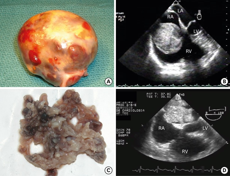
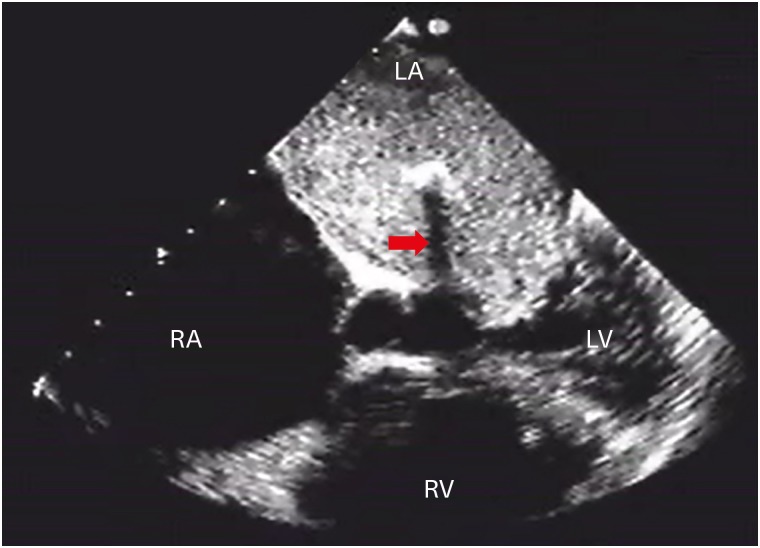
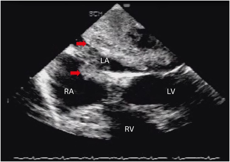
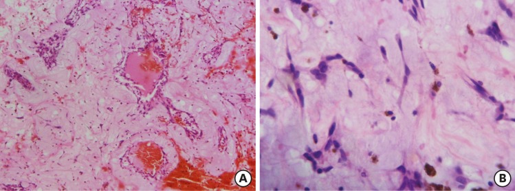
The patient's mean age was 53 ± 16 years (62.3% were women). The most common findings were dyspnea followed by embolic events. Most tumors were localized in the left atrium (77.4%), mainly in the fossa ovalis (63%). Mean size of the tumors was 4.76 x 3.50 cm. Tumors were generally mobile (88%) and went beyond the valve plane, causing mild mitral or tricuspid valve obstruction (58%) and dilation of the respective atrial chamber. Patients whose tumors were obstructive had higher pulmonary artery systolic pressures (50 vs 33 mmHg p < 0.01). According to the echocardiographic appearance 67% of tumors had a smooth surface and the remaining 32% had a villous surface. All patients with embolic manifestations had tumors with a villous surface.
Clinical presentation relates to the ultrasound characteristics of myxomas. Smooth tumors are larger, occur with obstructive symptoms, and benefit from an elective surgery, whereas villous myxomas entailed a high embolic risk and require prompt surgical treatment.
心脏黏液瘤是最常见的心脏肿瘤。尽管组织学上为良性,但在某些情况下,黏液瘤可能是致命的,这是由于其会损害心脏动力学以及具有血栓栓塞的可能性。本研究旨在评估心脏黏液瘤的临床表现及其与超声心动图特征的相关性,并描述手术治疗患者的围手术期结果和长期预后。
对53例在阿杰里奇医院接受手术的心脏黏液瘤患者进行前瞻性研究,从1993年至2013年对其进行临床随访和超声心动图检查。所有患者均接受了超声心动图检查。
患者的平均年龄为53±16岁(62.3%为女性)。最常见的表现是呼吸困难,其次是栓塞事件。大多数肿瘤位于左心房(77.4%),主要位于卵圆窝(63%)。肿瘤的平均大小为4.76×3.50厘米。肿瘤通常是可移动的(88%),并超出瓣膜平面,导致轻度二尖瓣或三尖瓣梗阻(58%)以及相应心房腔扩张。肿瘤有梗阻的患者肺动脉收缩压较高(50对33 mmHg,p<0.01)。根据超声心动图表现,67%的肿瘤表面光滑,其余32%的肿瘤表面呈绒毛状。所有有栓塞表现的患者其肿瘤表面均为绒毛状。
临床表现与黏液瘤的超声特征相关。表面光滑的肿瘤较大,出现梗阻症状,适合择期手术,而绒毛状黏液瘤有较高的栓塞风险,需要及时进行手术治疗。