Department of Pharmacology, University of Michigan Medical School, Ann Arbor, MI, United States.
Department of Pharmacology, University of Michigan Medical School, Ann Arbor, MI, United States; Departments of Physics and LSA Biophysics, University of Michigan, Ann Arbor, MI, United States.
Neurosci Lett. 2019 Apr 23;699:134-139. doi: 10.1016/j.neulet.2019.01.056. Epub 2019 Jan 31.
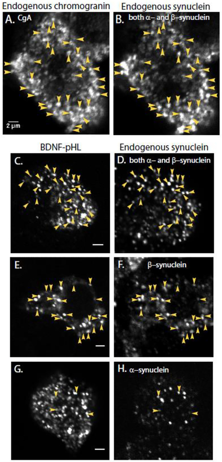
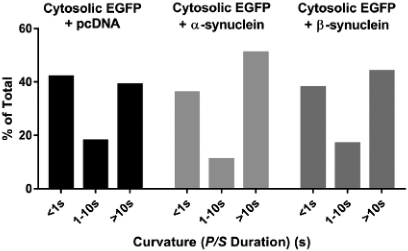
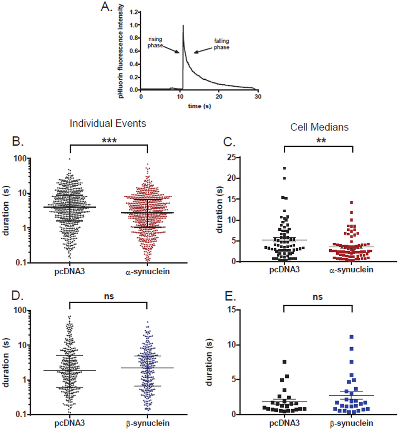
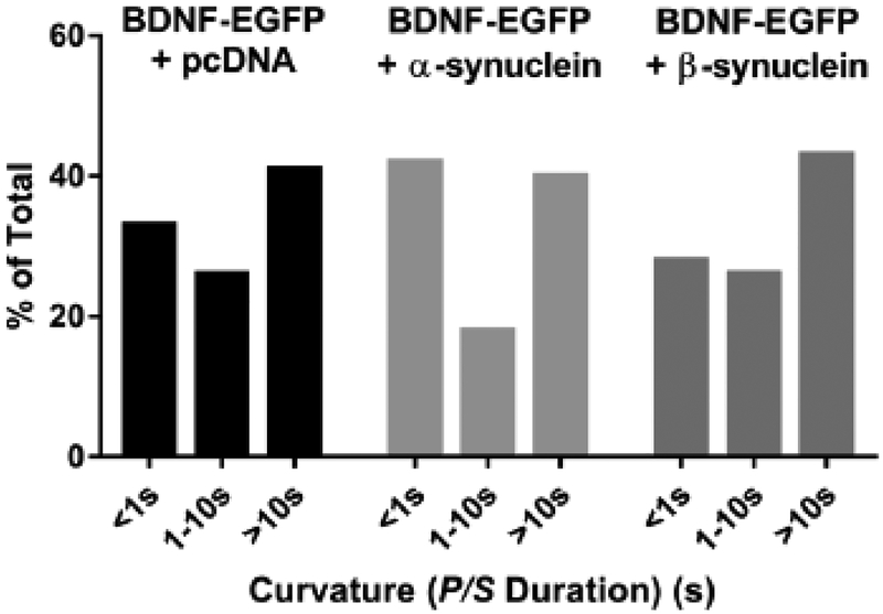
α-Synuclein is strongly implicated in the pathogenesis of Parkinson's disease as well as in other neurodegenerative diseases. However, its normal function in cells is not understood. The N-termini of α-, β-, and γ-synuclein contains six to seven 11-amino acid repeats that are predicted to form amphipathic helices. Membrane-binding and membrane-curving abilities of synuclein raise the possibility that synuclein could alter cellular processes that involve highly curved structures. In the present study we examined the localization of endogenous synuclein in bovine chromaffin cells by immunocytochemistry and its possible function to control protein discharge upon fusion of the granule with the plasma membrane by regulating the fusion pore. We found with quantitative immunocytochemistry that endogenous β-synuclein associates with secretory granules. Endogenous α-synuclein only rarely co-localizes with secretory granules. Overexpression of α-synuclein but not β-synuclein quickened the post- fusion discharge of BDNF-pHluorin by approxinately 30%. However, neither α- nor β-synuclein significantly altered curvature dynamics associated with fusion pore expansion that were measured by the combination of polarization and total internal reflection fluorescence microscopy (pTIRFM). Whatever the mechanism, the physiological significance of the small increased rate of post-fusion protein discharge caused by α-synuclein remains to be demonstrated, especially since endogenous β-, but not α-synuclein is the predominant synuclein isoform associated with chromaffin granules.
α-突触核蛋白强烈参与帕金森病以及其他神经退行性疾病的发病机制。然而,其在细胞中的正常功能尚不清楚。α-、β-和 γ-突触核蛋白的 N 端包含六个到七个 11 个氨基酸重复序列,预测其能形成两亲性螺旋。突触核蛋白的膜结合和膜弯曲能力提出了这样一种可能性,即突触核蛋白可能改变涉及高度弯曲结构的细胞过程。在本研究中,我们通过免疫细胞化学检查了内源性突触核蛋白在牛嗜铬细胞中的定位,以及通过调节融合孔来控制颗粒与质膜融合时蛋白质释放的可能功能。我们发现,通过定量免疫细胞化学,内源性β-突触核蛋白与分泌颗粒相关联。内源性 α-突触核蛋白很少与分泌颗粒共定位。α-突触核蛋白的过表达而不是β-突触核蛋白的过表达可使 BDNF-pHluorin 的融合后释放速度加快约 30%。然而,α-和β-突触核蛋白都没有显著改变融合孔扩张相关的曲率动力学,这是通过偏振和全内反射荧光显微镜(pTIRFM)的组合来测量的。无论机制如何,α-突触核蛋白引起的融合后蛋白质释放的小幅度增加的生理意义仍有待证明,特别是因为内源性β-突触核蛋白,而不是 α-突触核蛋白,是与嗜铬颗粒相关的主要突触核蛋白同工型。