Department of Biological Sciences, University of Southern California, Los Angeles, California, United States of America.
Department of Chemistry, University of Southern California, Los Angeles, California, United States of America.
PLoS Genet. 2019 Feb 4;15(2):e1007956. doi: 10.1371/journal.pgen.1007956. eCollection 2019 Feb.
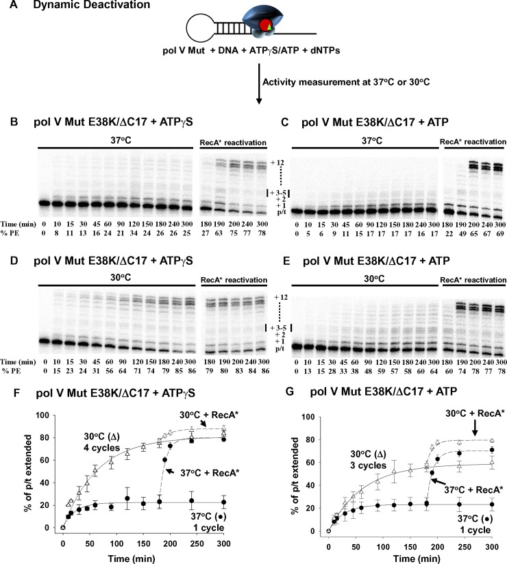
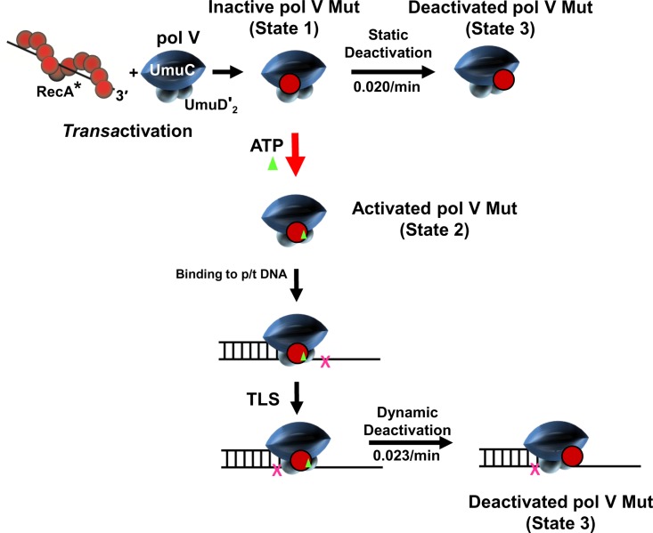
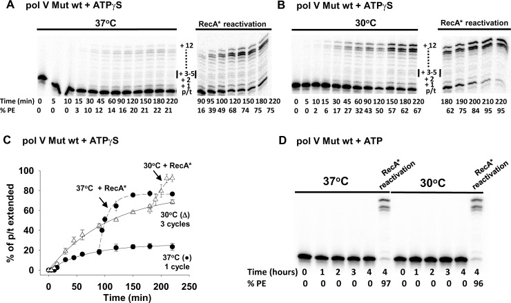
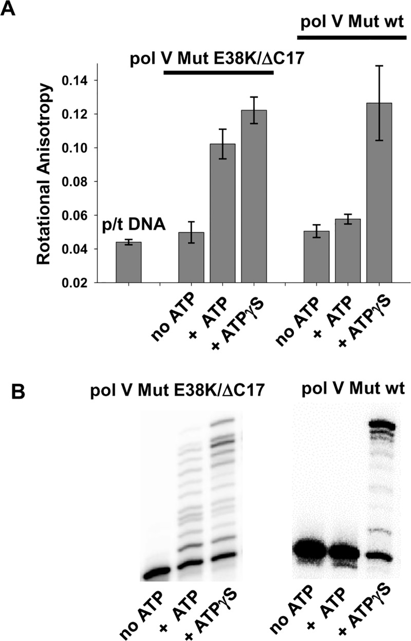
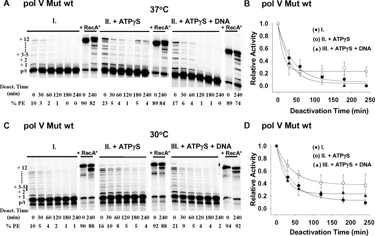
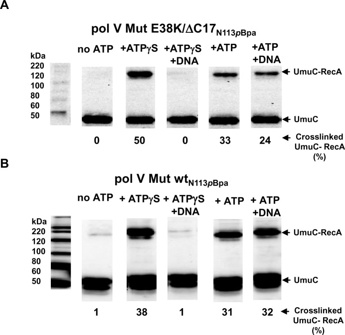
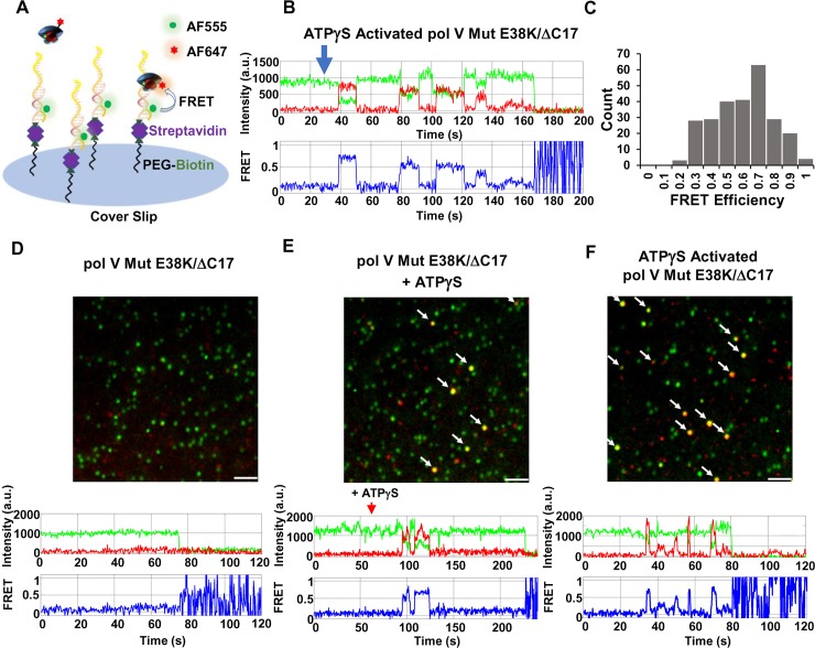
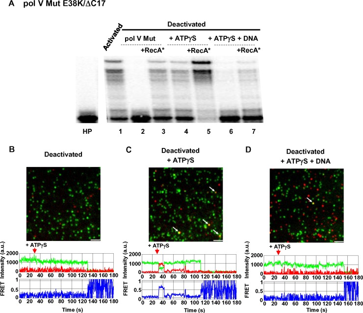
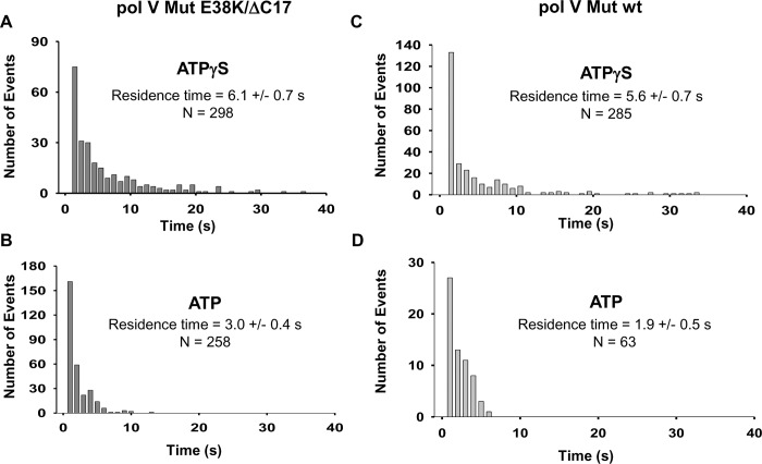
Mutagenic translesion DNA polymerase V (UmuD'2C) is induced as part of the DNA damage-induced SOS response in Escherichia coli, and is subjected to multiple levels of regulation. The UmuC subunit is sequestered on the cell membrane (spatial regulation) and enters the cytosol after forming a UmuD'2C complex, ~ 45 min post-SOS induction (temporal regulation). However, DNA binding and synthesis cannot occur until pol V interacts with a RecA nucleoprotein filament (RecA*) and ATP to form a mutasome complex, pol V Mut = UmuD'2C-RecA-ATP. The location of RecA relative to UmuC determines whether pol V Mut is catalytically on or off (conformational regulation). Here, we present three interrelated experiments to address the biochemical basis of conformational regulation. We first investigate dynamic deactivation during DNA synthesis and static deactivation in the absence of DNA synthesis. Single-molecule (sm) TIRF-FRET microscopy is then used to explore multiple aspects of pol V Mut dynamics. Binding of ATP/ATPγS triggers a conformational switch that reorients RecA relative to UmuC to activate pol V Mut. This process is required for polymerase-DNA binding and synthesis. Both dynamic and static deactivation processes are governed by temperature and time, in which on → off switching is "rapid" at 37°C (~ 1 to 1.5 h), "slow" at 30°C (~ 3 to 4 h) and does not require ATP hydrolysis. Pol V Mut retains RecA in activated and deactivated states, but binding to primer-template (p/t) DNA occurs only when activated. Studies are performed with two forms of the polymerase, pol V Mut-RecA wt, and the constitutively induced and hypermutagenic pol V Mut-RecA E38K/ΔC17. We discuss conformational regulation of pol V Mut, determined from biochemical analysis in vitro, in relation to the properties of pol V Mut in RecA wild-type and SOS constitutive genetic backgrounds in vivo.
突变型跨损伤 DNA 聚合酶 V(UmuD'2C)是大肠杆菌中 DNA 损伤诱导的 SOS 反应的一部分,受到多种水平的调控。UmuC 亚基被隔离在细胞膜上(空间调控),并在形成 UmuD'2C 复合物后进入细胞质,约在 SOS 诱导后 45 分钟(时间调控)。然而,只有当 pol V 与 RecA 核蛋白丝(RecA*)和 ATP 相互作用形成 mutasome 复合物,pol V Mut = UmuD'2C-RecA-ATP 时,才能进行 DNA 结合和合成。RecA 相对于 UmuC 的位置决定了 pol V Mut 是否具有催化活性(构象调控)。在这里,我们提出了三个相互关联的实验来解决构象调控的生化基础。我们首先研究了 DNA 合成过程中的动态失活和无 DNA 合成时的静态失活。然后,使用单分子(sm)TIRF-FRET 显微镜来探索 pol V Mut 动力学的多个方面。ATP/ATPγS 的结合触发构象转换,使 RecA 相对于 UmuC 重新取向,从而激活 pol V Mut。这个过程是聚合酶-DNA 结合和合成所必需的。动态失活和静态失活过程都受温度和时间的控制,在 37°C(1 到 1.5 小时)时,“快速”切换为 on→off,在 30°C(3 到 4 小时)时,“缓慢”切换为 on→off,并且不需要 ATP 水解。Pol V Mut 保持 RecA 在激活和失活状态,但只有在激活时才与引物-模板(p/t)DNA 结合。研究采用两种形式的聚合酶,pol V Mut-RecA wt 和组成型诱导和超突变的 pol V Mut-RecA E38K/ΔC17 进行。我们讨论了体外生化分析中确定的 pol V Mut 的构象调控,与体内 pol V Mut 在 RecA 野生型和 SOS 组成型遗传背景下的性质有关。