Zhang Michael, Radford Kennett D, Driscoll Mercedes, Purnomo Salsabila, Kim Jean, Choi Kwang H
Department of Psychiatry, Uniformed Services University, 4301 Jones Bridge Road, Bethesda, MD 20814, United States.
Center for the Study of Traumatic Stress, Uniformed Services University, 4301 Jones Bridge Road, Bethesda, MD 20814, United States.
IBRO Rep. 2019 Jan 16;6:87-94. doi: 10.1016/j.ibror.2019.01.006. eCollection 2019 Jun.
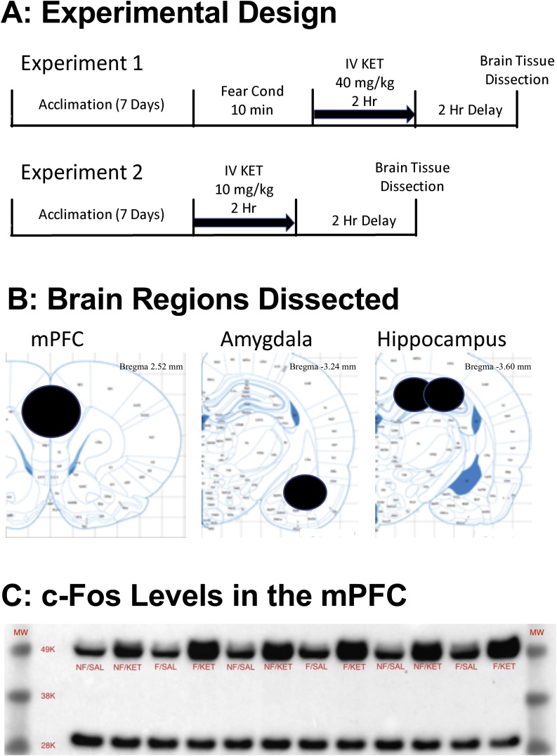
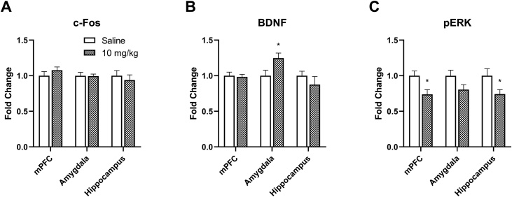
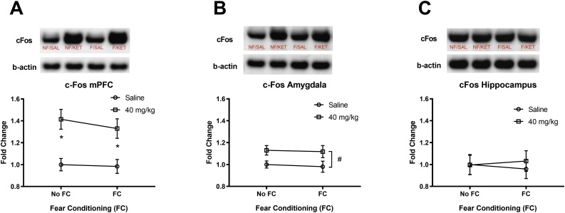
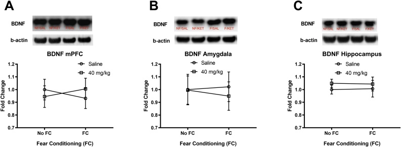
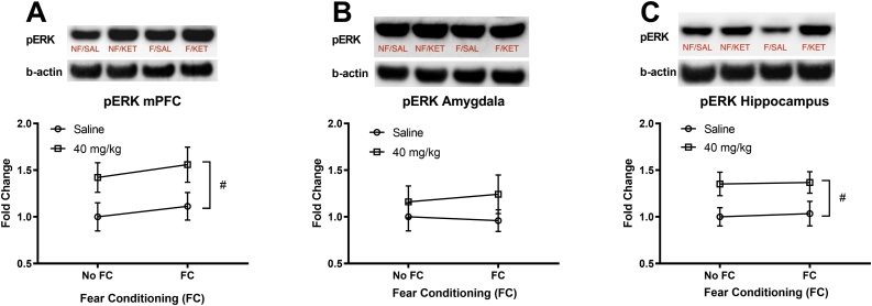
Ketamine, a multimodal dissociative anesthetic, is a powerful analgesic administered following trauma due to its hemodynamic and respiratory stability. However, ketamine can cause hallucination and dissociation which may adversely impact traumatic memory after an injury. The effects of ketamine on proteins implicated in neural plasticity are unclear due to different doses, routes, and timing of drug administration in previous studies. Here, we investigated the effects of a single intravenous (IV) ketamine infusion on protein levels in three brain regions of rats. Adult male Sprague-Dawley rats with indwelling IV catheters underwent an auditory fear conditioning (three pairings of tone and mild footshock 0.8 mA, 0.5 s) and received a high dose of IV ketamine (0 or 40 mg/kg/2 h) infusion (Experiment 1). In a follow-up study, animals received a low dose of IV ketamine (0 or 10 mg/kg/2 h) infusion (Experiment 2). Two hours after the infusion, brain tissue from the medial prefrontal cortex (mPFC), hippocampus, and amygdala were collected for western blot analyses. Protein levels of a transcription factor (c-Fos), brain-derived neurotrophic factor (BDNF), and phosphorylated extracellular signal-regulated kinase (pERK) were quantified in these regions. The 40 mg/kg ketamine infusion increased c-Fos levels in the mPFC and amygdala as well as pERK levels in the mPFC and hippocampus. The 10 mg/kg ketamine infusion increased BDNF levels in the amygdala, but decreased pERK levels in the mPFC and hippocampus. These findings suggest that a clinically relevant route of ketamine administration produces dose-dependent and brain region-specific effects on proteins involved in neuroplasticity.
氯胺酮是一种多模式解离麻醉剂,因其血流动力学和呼吸稳定性,在创伤后作为一种强效镇痛药使用。然而,氯胺酮会导致幻觉和解离,这可能会对受伤后的创伤记忆产生不利影响。由于先前研究中药物给药的剂量、途径和时间不同,氯胺酮对神经可塑性相关蛋白质的影响尚不清楚。在此,我们研究了单次静脉注射氯胺酮对大鼠三个脑区蛋白质水平的影响。成年雄性Sprague-Dawley大鼠留置静脉导管后进行听觉恐惧条件反射(音调与0.8毫安、0.5秒的轻度足部电击配对三次),并接受高剂量静脉注射氯胺酮(0或40毫克/千克/2小时)输注(实验1)。在后续研究中,动物接受低剂量静脉注射氯胺酮(0或10毫克/千克/2小时)输注(实验2)。输注两小时后,收集内侧前额叶皮质(mPFC)、海马体和杏仁核的脑组织进行蛋白质印迹分析。对这些区域中一种转录因子(c-Fos)、脑源性神经营养因子(BDNF)和磷酸化细胞外信号调节激酶(pERK)的蛋白质水平进行定量。40毫克/千克氯胺酮输注增加了mPFC和杏仁核中的c-Fos水平以及mPFC和海马体中的pERK水平。10毫克/千克氯胺酮输注增加了杏仁核中的BDNF水平,但降低了mPFC和海马体中的pERK水平。这些发现表明,氯胺酮的临床相关给药途径对参与神经可塑性的蛋白质产生剂量依赖性和脑区特异性影响。