Department of Biomedical Sciences, 2008 Vet Med Building, Iowa State University, Ames, IA, USA.
Department of Veterinary Diagnostic and Production Animal Medicine, 2203 Lloyd Veterinary Medical Center, Iowa State university, Ames, IA, USA.
Respir Res. 2019 Feb 6;20(1):27. doi: 10.1186/s12931-019-0992-3.
Animal production workers are persistently exposed to organic dust and can suffer from a variety of respiratory disease symptoms and annual decline in lung function. The role of high mobility group box-1 (HMGB1) in inflammatory airway diseases is emerging. Hence, we tested a hypothesis that organic dust exposure of airway epithelial cells induces nucleocytoplasmic translocation of HMGB1 and blocking this translocation dampens organic dust-induced lung inflammation.
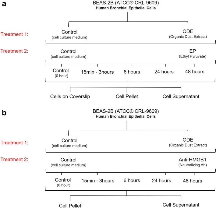
Rats were exposed to either ambient air or swine barn (8 h/day for either 1, 5, or 20 days) and lung tissues were processed for immunohistochemistry. Swine barn dust was collected and organic dust extract (ODE) was prepared and sterilized. Human airway epithelial cell line (BEAS-2B) was exposed to either media or organic dust extract followed by treatment with media or ethyl pyruvate (EP) or anti-HMGB1 antibody. Immunoblotting, ELISA and other assays were performed at 0 (control), 6, 24 and 48 h. Data (as mean ± SEM) was analyzed using one or two-way ANOVA followed by Bonferroni's post hoc comparison test. A p value of less than 0.05 was considered significant.
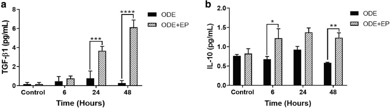
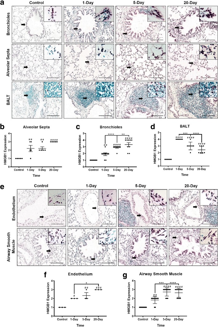
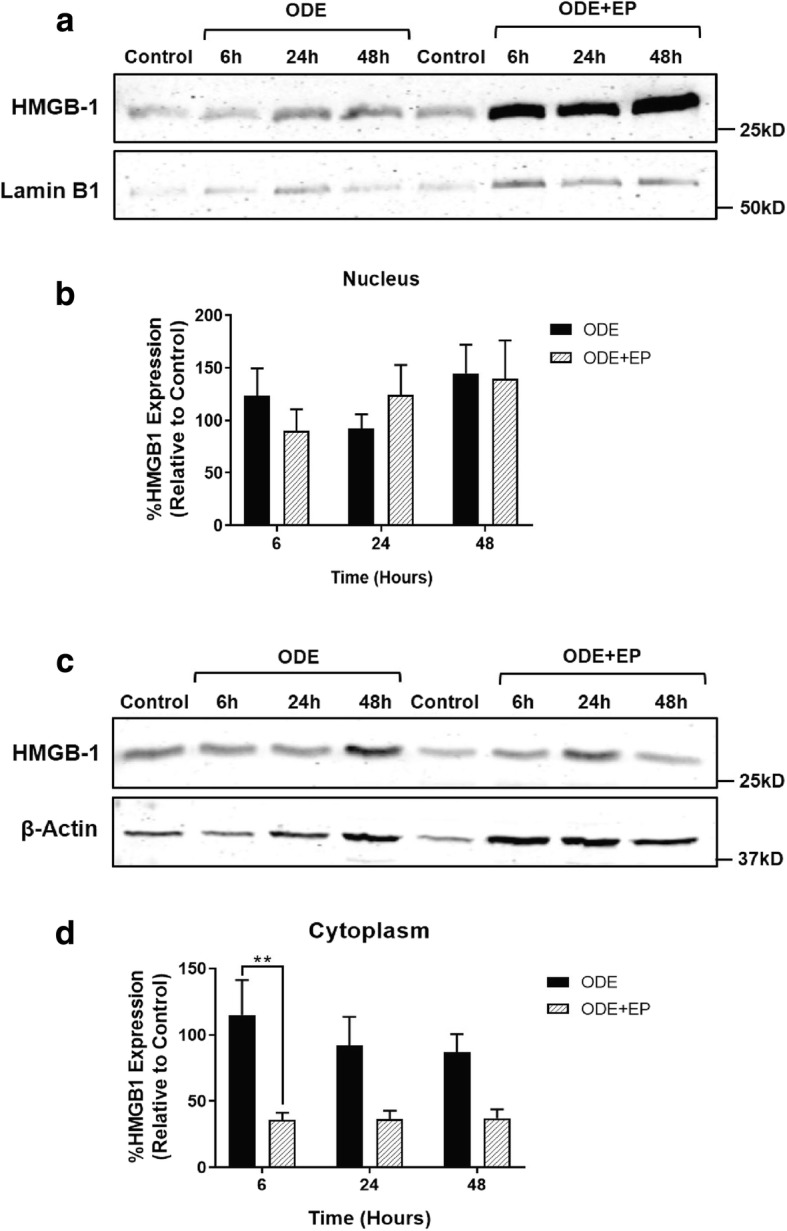
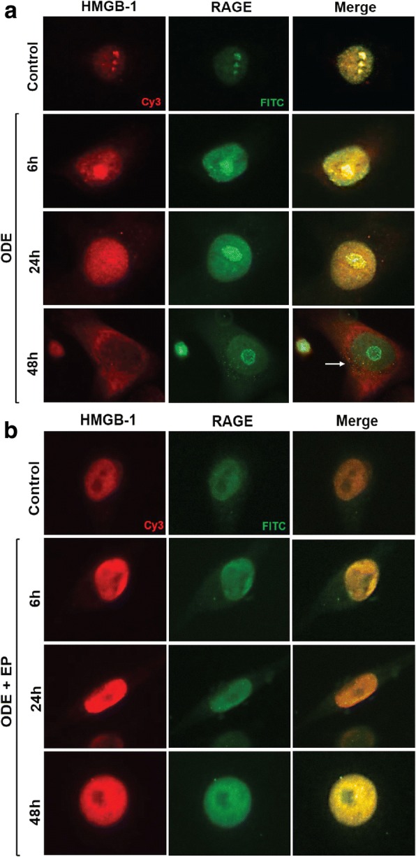
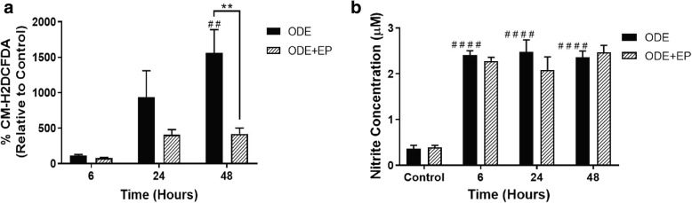
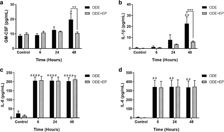
Compared to controls, barn exposed rats showed an increase in the expression of HMGB1 in the lungs. Compared to controls, ODE exposed BEAS-2B cells showed nucleocytoplasmic translocation of HMGB1, co-localization of HMGB1 and RAGE, reactive species and pro-inflammatory cytokine production. EP treatment reduced the ODE induced nucleocytoplasmic translocation of HMGB1, HMGB1 expression in the cytoplasmic fraction, GM-CSF and IL-1β production and augmented the production of TGF-β1 and IL-10. Anti-HMGB1 treatment reduced ODE-induced NF-κB p65 expression, IL-6, ROS and RNS but augmented TGF-β1 and IL-10 levels.
HMGB1-RAGE signaling is an attractive target to abrogate OD-induced lung inflammation.
动物生产工人持续暴露于有机粉尘中,可能会遭受各种呼吸道疾病症状,并出现肺功能逐年下降。高迁移率族蛋白 B1(HMGB1)在炎症性气道疾病中的作用正在显现。因此,我们提出了一个假设,即气道上皮细胞暴露于有机粉尘会诱导 HMGB1 的核质转位,阻断这种转位可减轻有机粉尘引起的肺部炎症。
将大鼠暴露于环境空气或猪舍(每天 8 小时,分别暴露 1、5 或 20 天),并处理肺组织进行免疫组织化学分析。收集猪舍粉尘,制备并消毒有机粉尘提取物(ODE)。将人气道上皮细胞系(BEAS-2B)暴露于培养基或有机粉尘提取物中,然后用培养基或乙基丙酮酸(EP)或抗 HMGB1 抗体处理。在 0(对照)、6、24 和 48 小时时进行免疫印迹、ELISA 和其他检测。使用单因素或双因素方差分析,然后进行 Bonferroni 事后比较检验分析数据(作为均值±SEM)。p 值小于 0.05 被认为具有统计学意义。
与对照组相比,暴露于猪舍的大鼠肺部 HMGB1 的表达增加。与对照组相比,ODE 暴露的 BEAS-2B 细胞显示 HMGB1 发生核质转位,HMGB1 与 RAGE 共定位,活性氧和促炎细胞因子产生。EP 处理减少了 ODE 诱导的 HMGB1 核质转位、细胞质部分 HMGB1 表达、GM-CSF 和 IL-1β 产生,并增加了 TGF-β1 和 IL-10 的产生。抗 HMGB1 治疗减少了 ODE 诱导的 NF-κB p65 表达、IL-6、ROS 和 RNS,但增加了 TGF-β1 和 IL-10 水平。
HMGB1-RAGE 信号是阻断 OD 诱导的肺部炎症的一个有吸引力的靶点。